. 2014 Mar 1;31(5):476-86.
doi: 10.1089/neu.2013.3135. Epub 2013 Dec 19.

Neuroprotective efficacy of a proneurogenic compound after traumatic brain injury

Affiliations

Neuroprotective efficacy of a proneurogenic compound after traumatic brain injury

Meghan O Blaya et al. J Neurotrauma. .

Abstract

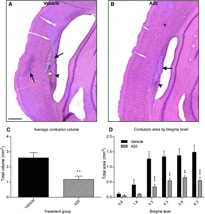
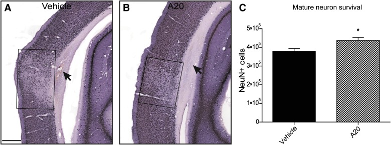
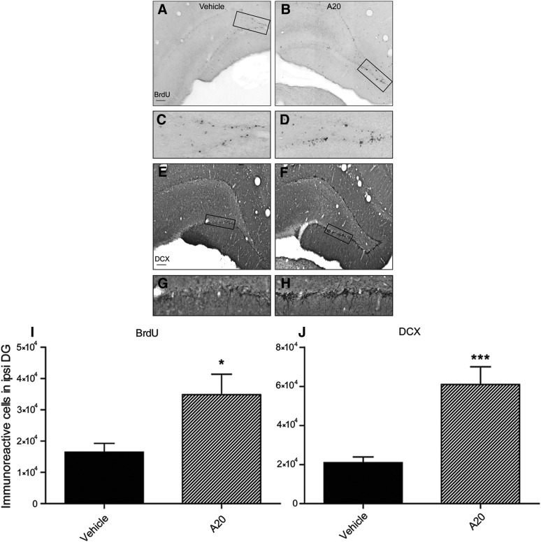
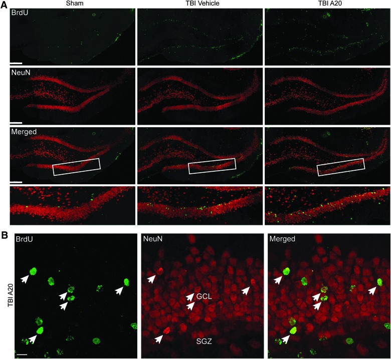
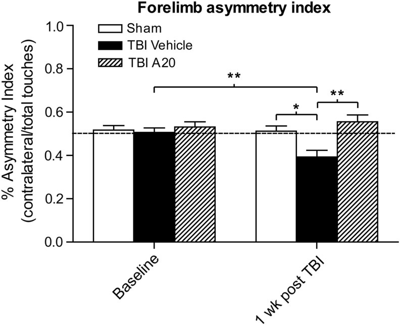
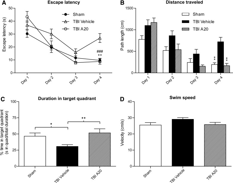
Traumatic brain injury (TBI) is characterized by histopathological damage and long-term sensorimotor and cognitive dysfunction. Recent studies have reported the discovery of the P7C3 class of aminopropyl carbazole agents with potent neuroprotective properties for both newborn neural precursor cells in the adult hippocampus and mature neurons in other regions of the central nervous system. This study tested, for the first time, whether the highly active P7C3-A20 compound would be neuroprotective, promote hippocampal neurogenesis, and improve functional outcomes after experimental TBI. Sprague-Dawley rats subjected to moderate fluid percussion brain injury were evaluated for quantitative immunohistochemical and behavioral changes after trauma. P7C3-A20 (10 mg/kg) or vehicle was initiated intraperitoneally 30 min postsurgery and twice per day every day thereafter for 7 days. Administration of P7C3-A20 significantly reduced overall contusion volume, preserved vulnerable anti-neuronal nuclei (NeuN)-positive pericontusional cortical neurons, and improved sensorimotor function 1 week after trauma. P7C3-A20 treatment also significantly increased both bromodeoxyuridine (BrdU)- and doublecortin (DCX)-positive cells within the subgranular zone of the ipsilateral dentate gyrus 1 week after TBI. Five weeks after TBI, animals treated with P7C3-A20 showed significantly increased BrdU/NeuN double-labeled neurons and improved cognitive function in the Morris water maze, compared to TBI-control animals. These results suggest that P7C3-A20 is neuroprotective and promotes endogenous reparative strategies after TBI. We propose that the chemical scaffold represented by P7C3-A20 provides a basis for optimizing and advancing new pharmacological agents for protecting patients against the early and chronic consequences of TBI.


Figures

<b>FIG. 1.</b>
FIG. 1.
Administration of A20 significantly decreased contusion volumes 1 week post-TBI. (A and B) Representative images showing contusions at gray/white matter interface in vehicle and A20 animals, respectively. Black arrows show contusions at ipsilateral external capsule and parietal cortex; arrowheads indicate areas of intracerebral hemorrhage. (C) Contusions were contoured and volumetric analyses were conducted at six bregma levels stained with H&E. A20 animals exhibited greater than a 2-fold decrease in average contusion volumes, compared to vehicle control animals (Student's t-test: p=0.0025; t=3.504). (D) A20 exerted significant cytoprotective effects at four of six bregma levels (epicenter of injury, −3.8 mm). Two-way repeated-measures ANOVA established significance for bregma level (p<0.0001), treatment (p=0.0012), and treatment×bregma level interaction (p=0.0002). Bonferroni's post-hoc analysis showed significant differences in contusion area at the four most posterior bregma levels evaluated (**p<0.01; ***p<0.001; scale bar, 500 μm). Data are expressed as mean±standard error of the mean; n=20. TBI, traumatic brain injury; H&E, hematoxylin and eosin; ANOVA, analysis of variance.
<b>FIG. 2.</b>
FIG. 2.
A20 administration was neuroprotective in vulnerable ipsilateral cortical regions 1 wk post TBI. (A and B) Representative NeuN-stained images of vehicle- (A) and A20-treated (B) animals. Selective neuronal loss was observed in the cortical region overlying the contusion site. Treatment with A20 significantly reduced NeuN-positive cell loss. Boxes in A, B enclose the areas of neuronal loss. Arrows indicate subcortical lesion. (C) Quantification of NeuN-positive cells between treatment groups revealed a significant neuroprotective effect of A20 1 wk post injury (Student's t-test: p=0.015, t=2.695; *p<0.05; scale bar, 500 μm); n=20. TBI, traumatic brain injury; NeuN, anti-neuronal nuclei.
<b>FIG. 3.</b>
FIG. 3.
Administration of A20 increased progenitor cell and immature neuron proliferation 1 week post-TBI. Representative images showing BrdU- (A–D) and DCX-immunoreactive cells (E–H) in TBI vehicle- (left) and A20- (right) treated groups. Images in (C and D) and (G and H) are magnified regions of boxed areas in (A), (B), (E), and (F). (I and J) Quantification of BrdU- and DCX- positive cells. A20 administration significantly increased both BrdU- and DCX-positive cells, indicative of early stage neurogenesis (Student's t-test: BrdU: p=0.0163, t=2.665; DCX: p=0.0006, t=4.174; *p<0.05; ***p<0.001; scale bars, 200 μm); n=20. TBI, traumatic brain injury; BrdU, bromodeoxyuridine; DCX, doublecortin.
<b>FIG. 4.</b>
FIG. 4.
Confocal micrographs showing BrdU- and NeuN-immunoreactive cells within the DG 5 weeks after TBI. (A) Representative fluorescent images showing cells that colabeled for BrdU and NeuN in the ipsilateral DG (scale bars, 200 μm). Boxed regions outline the areas of higher magnification shown in bottom row of (A). (B) Representative micrographs from a TBI-A20 animal showing several cells coexpressing markers for BrdU and NeuN (arrows) in the ipsilateral DG. Double-labeled cells in A20 animals appeared to migrate out of the SGZ into more superficial layers of the DG, consistent with neuronal maturation profiles (GCL, granular cell layer; SGZ, subgranular zone; scale bar, 10 μm). BrdU, bromodeoxyuridine; NeuN, anti-neuronal nuclei; DG, dentate gyrus; TBI, traumatic brain injury.
<b>FIG. 5.</b>
FIG. 5.
Quantitative assessment of hippocampal neurogenesis 5 weeks after TBI. A20-treated TBI animals demonstrated significant increases in numbers of BrdU/NeuN-positive cells, compared to vehicle-treated and sham-operated controls. TBI-vehicle controls had significantly more double-labeled cells than sham-operated animals. One-way analysis of variance showed significance (p<0.0001, F=46.16); Bonferroni's multiple comparison test showed significant differences among groups (***p<0.001; ****p<0.0001); n=27. BrdU, bromodeoxyuridine; NeuN, anti-neuronal nuclei; TBI, traumatic brain injury.
<b>FIG. 6.</b>
FIG. 6.
A20 protected from contralateral forelimb deficits 1 week post-TBI. An asymmetry index of less than 50% indicates a contralateral deficit. All animal groups performed similarly presurgery. One week after TBI, vehicle-treated controls exhibited a significant contralateral forelimb deficit, compared to their baseline time point (Student's t-test: p=0.0095; t=3.005). Contralateral forelimb usage in the TBI-vehicle control group 1 week postinjury was significantly decreased, compared to sham-uninjured animals, consistent with this injury model. In contrast, TBI animals treated with A20 showed no signs of a contralateral deficit and performed similar to sham-operated animals and significantly better than TBI-vehicle controls (one-way analysis of variance 1-week time point: p=0.0022, F=7.996; Bonferroni's multiple comparison test: sham vs. TBI-vehicle: *p<0.05; TBI-vehicle vs. TBI-A20: **p<0.01); n=27. TBI, traumatic brain injury.
<b>FIG. 7.</b>
FIG. 7.
A20 administration significantly improved Morris water maze performance 4 weeks after TBI. (A) Post-hoc analysis revealed that TBI-A20 and sham animals had significantly reduced escape latencies on the hidden platform task, relative to TBI-vehicle controls, on the fourth day of testing (Bonferroni's multiple comparison test: TBI-A20 vs. TBI-vehicle, **p<0.01; sham vs. TBI-vehicle, ###p<0.001). Two-way repeated measures analysis of variance (ANOVA) was significant for trial day (p<0.0001), treatment (p=0.007), and treatment×trial day interaction (p=0.0005). (B) By day 4, sham and TBI-A20 animals exhibited significantly shorter path lengths to reach the platform, compared to TBI-vehicle controls (Bonferroni's multiple comparison test: **p<0.01). Two-way repeated-measures ANOVA was significant for trial day (p<0.0001), treatment (p=0.0013), and treatment×trial day interaction (p<0.016). (C) On day 4, sham and TBI-A20 animals spent a greater percentage of time in the target quadrant, compared to TBI-vehicle control animals (Bonferroni's multiple comparison test: *p<0.05; **p<0.01). Two-way repeated-measures ANOVA showed significance for trial day (p<0.0001) and treatment (0.0168), but not for treatment×trial day interaction (p=0.131). (D) There were no significant differences in swim speeds among groups, suggesting that the injury did not affect swimming skills (one-way ANOVA: p=0.129); n=27. TBI, traumatic brain injury.

References

    1. Langlois J.A., and Rutland-Brown W. (2005) Traumatic Brain Injury in the United States: The Future of Registries and Data Systems. Centers for Disease Control and Prevention, National Center for Injury Prevention and Control: Atlanta, GA
    1. Bramlett H.M., and Dietrich W.D. (2007). Progressive damage after brain and spinal cord injury: pathomechanisms and treatment strategies. Prog. Brain Res. 161, 125–141 - PubMed
    1. Lowenstein D.H., Thomas M.J., Smith D.H., and McIntosh T.K. (1992). Selective vulnerability of dentate hilar neurons following traumatic brain injury: a potential mechanistic link between head trauma and disorders of the hippocampus. J. Neurosci. 12, 4846–4853 - PMC - PubMed
    1. Kotapka M.J., Gennarelli T.A., Graham D.I., Adams J.H., Thibault L.E., Ross D.T., and Ford I. (1991). Selective vulnerability of hippocampal neurons in acceleration-induced experimental head injury. J. Neurotrauma 8, 247–258 - PubMed
    1. Bramlett H.M., Green E.J., and Dietrich W.D. (1997). Hippocampally dependent and independent chronic spatial navigational deficits following parasagittal fluid percussion brain injury in the rat. Brain Res. 762, 195–202 - PubMed

Publication types

MeSH terms