. 2013 Sep 27;45(9):e44.
doi: 10.1038/emm.2013.87.

Constitutive stabilization of hypoxia-inducible factor alpha selectively promotes the self-renewal of mesenchymal progenitors and maintains mesenchymal stromal cells in an undifferentiated state

Affiliations

Constitutive stabilization of hypoxia-inducible factor alpha selectively promotes the self-renewal of mesenchymal progenitors and maintains mesenchymal stromal cells in an undifferentiated state

In-Ho Park et al. Exp Mol Med. .

Abstract

With the increasing use of culture-expanded mesenchymal stromal cells (MSCs) for cell therapies, factors that regulate the cellular characteristics of MSCs have been of major interest. Oxygen concentration has been shown to influence the functions of MSCs, as well as other normal and malignant stem cells. However, the underlying mechanisms of hypoxic responses and the precise role of hypoxia-inducible factor-1α (Hif-1α), the master regulatory protein of hypoxia, in MSCs remain unclear, due to the limited span of Hif-1α stabilization and the complex network of hypoxic responses. In this study, to further define the significance of Hif-1α in MSC function during their self-renewal and terminal differentiation, we established adult bone marrow (BM)-derived MSCs that are able to sustain high level expression of ubiquitin-resistant Hif-1α during such long-term biological processes. Using this model, we show that the stabilization of Hif-1α proteins exerts a selective influence on colony-forming mesenchymal progenitors promoting their self-renewal and proliferation, without affecting the proliferation of the MSC mass population. Moreover, Hif-1α stabilization in MSCs led to the induction of pluripotent genes (oct-4 and klf-4) and the inhibition of their terminal differentiation into osteogenic and adipogenic lineages. These results provide insights into the previously unrecognized roles of Hif-1α proteins in maintaining the primitive state of primary MSCs and on the cellular heterogeneities in hypoxic responses among MSC populations.


Figures
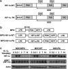
Figure 1
Figure 1
Kinetics of protein stabilization for various forms of Hif-1α. (a) Schematic structure of wild-type (WT) and ubiquitin-resistant form of Hif-1 α (PA). bHLH; basic helix-loop-helix domain, PAS; Per/Arnt/Sim, ODD; oxygen-dependent degradation domain, TAD; transactivation domain. The proline residues (P402, p564) in ODD were substituted for Ala residues in the Hif-1α-PA mutant. (b) Structures of retroviral vectors WT or PA mutant form of Hif-1α along with green fluorescent protein (GFP). (c) Comparisons for kinetics of Hif-1 α and Hif-1β (ARNT) protein upon exposure to hypoxia (1% O2). Shown are representative western blots for each indicated protein in MSCs during incubation under hypoxic conditions.
Figure 2
Figure 2
Effects of hypoxia and Hif-1α stabilization on the growth of MSCs in mass population. (a) BrdU incorporation analysis of MSCs under normoxic or hypoxic conditions. Cells were added with 10 μM of BrdU in the culture medium for 2 h. MSCs were then stained with antibody against BrdU and propidium iodide (PI) for flow cytometric analysis. Shown are the representative plots and percentage of cells in each cell cycle fraction. (b) Effects of hypoxia on cell cycle inhibitory proteins in MSCs. MSCs were cultured under 21 or 1% O2 for 2 days and analyzed for protein levels of each indicated gene by using antibody specific to p21 or p27. (c) MSCs were maintained under hypoxic (1% O2) or normoxic (21% O2) for at least two passages and plated in the dish (5 × 10⁴/100 mm dish density) at equal density. Eight days after plating, the cell numbers were counted. Shown are the mean±s.e.m. values from three experiments. (d) Doubling times were calculated as t/n, where t is the duration of culture and n is the number of population doublings calculated by using the formula n=log(NH−NI)/log2 (where NI is the number of cells originally plated and NH the number of cells harvested at the time of counting). Shown are the mean±s.e.m. values from three experiments. APC, allophycocyanin.
Figure 3
Figure 3
(a) Effects of hypoxia and Hif-1α stabilization during expansion culture on the self-renewal of colony-forming mesenchymal progenitor cells. (Upper) Illustration of the experimental scheme. Control (MSC/MPG) or Hif-1α-PA-transduced MSCs (MSC/PA) were maintained under normoxic (21%) or hypoxic (1%) conditions for two passages (7 days), equal numbers of each group of MSCs were then plated for colonization under normoxia. (Middle) Representative morphology of colony formation for each group of MSCs. (Lower) Number of CFU-F formed from plating 1000 cells of each group of MSCs in the 100-mm dish. Shown are the mean±s.e.m. values from four independent experiments. *P<0.05. (b) Effects of hypoxia and Hif-1α stabilization on the colonization process of MSCs. (Upper) Illustration of the experimental scheme. Control (MSC/MPG) or Hif-1α-PA-transduced MSCs (MSC/PA) were maintained under normoxic (21%) or hypoxic (1%) conditions for two passages (7 days), then equal number (1000/well) of each group of MSCs were plated under normoxic (21%) or hypoxic (1%) conditions for colonization of mesenchymal progenitors. (Middle) Representative morphology of colony formation for each group of MSCs. (Lower) Number of CFU-F formed from plating 1000 cells of each group of MSCs in a100-mm dish. Shown are the mean±s.e.m. values from four independent experiments. *P<0.05.
Figure 4
Figure 4
Effects of Hif-1α stabilization on the expression of pluripotent genes in MSC. Control (MSC/MPG) or Hif-1α-PA-transduced MSCs (MSC/PA) were exposed to 1% hypoxia and expression of each indicated gene was analyzed by reverse transcriptase-PCR. GAPDH, glyceraldehyde 3-phosphate dehydrogenase.
Figure 5
Figure 5
Effects of hypoxia and Hif-1α stabilization on the osteogenic differentiation of MSCs. (a, b) Control (MSC/MPG) or Hif-1α-PA-transduced MSCs (MSC/PA) were subjected to osteogenic differentiation by chemically defined medium under normoxic or hypoxic (1%) conditions. At 14 days after differentiation, the cells were examined by Alizarin Red S staining and quantification of mineralized nodules as determined by absorbance at 550 nm after dye elution. Shown are representative morphology and mean±s.e.m. values from four independent experiments. *P<0.05. (c) Expression of osteogenic gene expression during differentiation was analyzed by reverse transcriptase-PCR. (d) Stabilization of TAZ during osteogenic differentiation of each group of MSCs were compared with each group of MSCs during normoxia and hypoxia (1% O2). GAPDH, glyceraldehyde 3-phosphate dehydrogenase; OC, osteocalcin; OP, osteopontin.
Figure 6
Figure 6
Effects of hypoxia and Hif-1α on the adipogenic differentiation of MSCs. Each group of MSCs were subjected to adipogenic differentiation by chemically defined media under normoxic or hypoxic conditions. (a, b) After 7 days in their induction media, MSCs were stained with Oil-Red O and lipid accumulation was quantified by dye extraction and spectrophotometry at 502 nm. Shown are the mean±s.e.m. values from four independent experiments. *P<0.05. (c) Expression of peroxisome proliferator-activated receptor-γ (PPARγ) during adipogenic differentiation was analyzed by reverse transcriptase-PCR. GAPDH, glyceraldehyde 3-phosphate dehydrogenase.
Figure 7
Figure 7
Effects of hypoxia and Hif-1α on the neuronal differentiation of MSCs. Each indicated group of MSCs were subjected to neuronal differentiation by chemically defined media under normoxic or hypoxic conditions. Twenty-four hours after induction under normoxic or hypoxic conditions, MSCs were examined by (a) morphology and the expression of each indicated neuron-specific genes was determined by (b) western blot analysis. (c) Original current recordings in normoxic or hypoxic cells. Whole-cell current was elicited by a 50-ms depolarizing pulse to +40 mV from a holding potential of −120 mV at 10-s intervals.

References

    1. Keating A. Mesenchymal stromal cells. Curr Opin Hematol. 2006;13:419–425. - PMC - PubMed
    1. Pittenger MF, Mackay AM, Beck SC, Jaiswal RK, Douglas R, Mosca JD, et al. Multilineage potential of adult human mesenchymal stem cells. Science. 1999;284:143–147. - PubMed
    1. Prockop DJ. Marrow stromal cells as stem cells for nonhematopoietic tissues. Science. 1997;276:71–74. - PubMed
    1. Caplan AI, Dennis JE. Mesenchymal stem cells as trophic mediators. J Cell Biochem. 2006;98:1076–1084. - PubMed
    1. Giordano A, Galderisi U, Marino IR. From the laboratory bench to the patient's bedside: an update on clinical trials with mesenchymal stem cells. J Cell Physiol. 2007;211:27–35. - PubMed

Publication types

MeSH terms

Substances

LinkOut - more resources