. 2013 Sep 27:3:2786.
doi: 10.1038/srep02786.

Autologous transplantation of GDNF-expressing mesenchymal stem cells protects against MPTP-induced damage in cynomolgus monkeys

Affiliations

Autologous transplantation of GDNF-expressing mesenchymal stem cells protects against MPTP-induced damage in cynomolgus monkeys

Zhenhua Ren et al. Sci Rep. .

Abstract

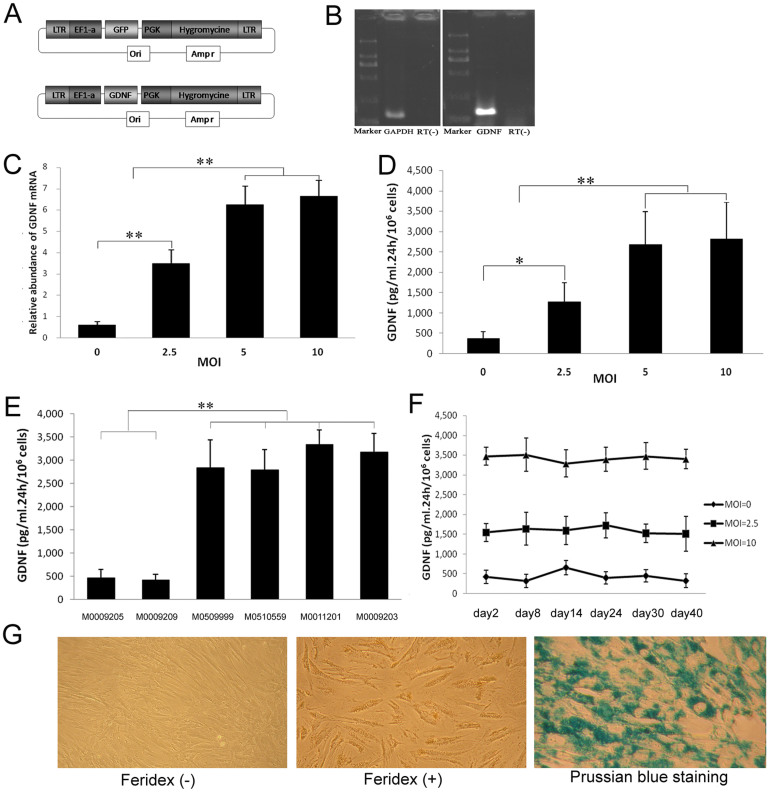
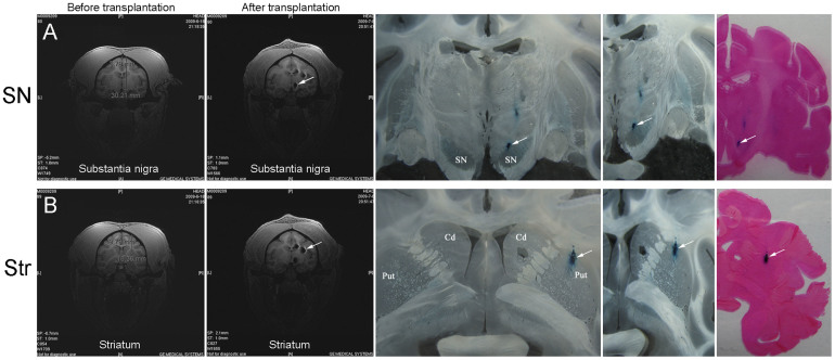
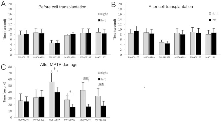
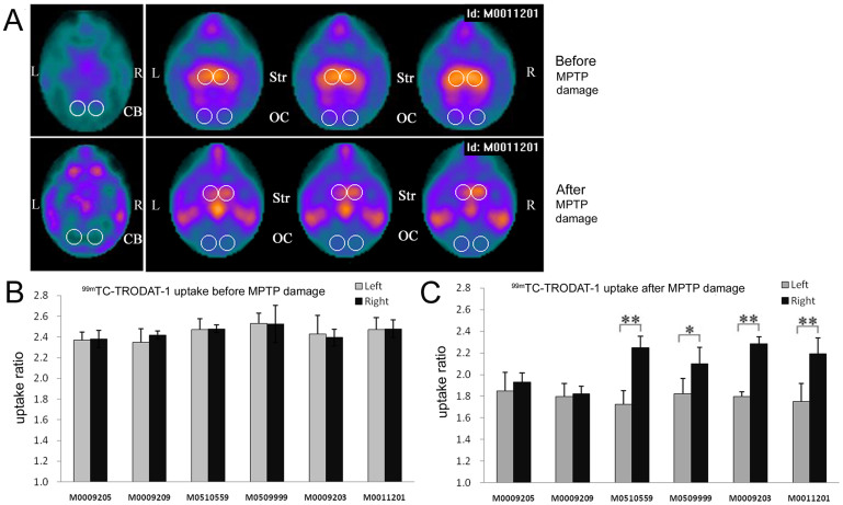
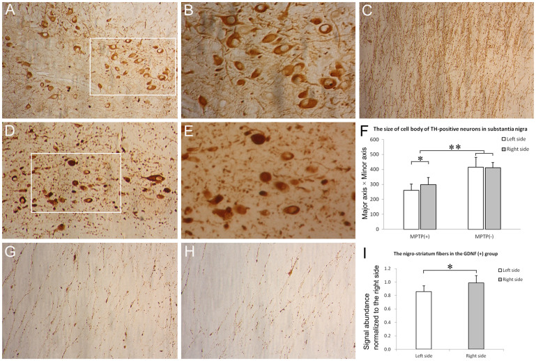
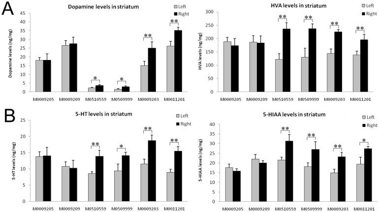
Glial cell-derived neurotrophic factor (GDNF) has shown beneficial effects in models of Parkinson's disease. The mild results observed in the double-blind clinical trial by intraputamenal infusion of recombinant GDNF proteins warrant a search for alternative delivery methods. In this study, we investigated the function of autologous mesenchymal stem cells (MSCs) expressing GDNF (GDNF-MSCs) for protection against MPTP-induced injury in cynomolgus monkeys. MSCs were obtained from the bone marrow of individual monkeys and gene-modified to express GDNF. Following unilateral engraftment of GDNF-MSCs into the striatum and substantia nigra, the animals were challenged with MPTP to induce a stable systemic Parkinsonian state. The motor functions were spared in the contralateral limbs of monkeys receiving GDNF-MSCs, but not in those receiving MSCs alone. In the striatum of the grafted hemisphere, dopamine levels were higher and dopamine uptake was enhanced. The results suggest that autologous MSCs may be a safe vehicle to deliver GDNF for enhancing nigro-striatum functions.


Figures

Figure 1
Figure 1. Culture and characterization of bone marrow mesenchymal stem cells.
(A) Schematic diagram of MSC isolation from bone marrow by using Ficoll- Paque™ Plus. (B) Primary culture of MSCs at day 8 (200×). (C) Cynomolgus monkey MSCs showed a normal karyotype of 42, XY. (D) Surface marker expression on MSCs at passage 3 analyzed by flow cytometry. Unfilled lines represent isotype controls. (E) In vitro differentiation of MSCs into adipocytes (Oil O Red staining, upper left), chondrocytes (Safranin O staining, upper middle) and osteocytes (von Kossa Staining, upper right). Staining of undifferentiated MSCs served as controls (lower panels).
Figure 2
Figure 2. Lentivirus infection and Feridex-labeling of MSCs.
(A) Schematic diagram of vector construction. (B) Expression of GDNF mRNA in MSCs after lentivirus infection. (C) Relative abundance of GDNF mRNA analyzed by real-time PCR in GDNF-MSCs at various MOIs. (D) GDNF secretion analyzed by ELISA in GDNF-MSCs at various MOIs. (E) Compared to the two non-infected MSC lines, GDNF secretion was significantly higher in MSCs infected with lenti-GDNF at MOI 10. (F) Stable expression of GDNF for 40 days. (G) Feridex-labeled MSCs appeared yellowish under bring field microscopy and could be visualized by Prussian blue staining. (C–E) *P < 0.05, **P < 0.01).
Figure 3
Figure 3. Tracking of transplanted cells.
(A) Transplantation into substantia nigra. MRI images, Prussian blue staining, and eosin staining all confirmed that the cells had been correctly targeted at the substantia nigra. (B) Transplantation into striatum. MRI images, Prussian blue staining, and eosin staining confirmed that the cells had been correctly deposited at the striatum. SN, substantia nigra; Str, striatum.
Figure 4
Figure 4. Motor functions are protected by GDNF-MSCs.
The left and right upper limb motor performance was recorded and the time spent to take the food treat was analyzed. (A) Before cell transplantation; (B) After transplantation but before MPTP treatment; (C) After transplantation and MPTP treatment. No difference was noticed in motor functions after transplantation. However, following MPTP treatment, all the 6 monkeys spent more time to get the food. All the 4 monkeys that had received GDNF-MSCs in the right hemisphere showed faster movement in the left upper limb (C). *P < 0.05, **P < 0.01.
Figure 5
Figure 5. 99mTc-TRODAT-1 uptake analyzed by SPECT imaging.
(A) Representative SPECT images for one monkey before (upper panels) and after (lower panels) MPTP treatment. The signal intensities in striatum were reduced after MPTP treatment. However, in the MPTP-treated animals, the right striatum showed higher signal intensities compared with the left striatum. The circled areas indicate the regions of interest. CB, cerebellum; OC, occipital cortex; Str, striatum. (B and C) The ratio of signal intensities in striatum relative to those in cerebellum and occipital cortex was measured (Str/[(CB + OC)/2]) before (B) and after (C) MPTP treatment. *P < 0.05, **P < 0.01.
Figure 6
Figure 6. Immunohistochemical analysis of dopaminergic system.
Compared with naive controls (A and B), the number of TH-positive neurons in the substantia nigra pars compacta was dramatically reduced in all MPTP-treated monkeys (D and E). (B) and (E) represent the magnified views of the insets in (A) and (D), respectively. However, no significant difference existed in the number of TH+ neurons in the left versus right half of substantia nigra either in the GDNF (+) or GDNF (-) group after MPTP treatment. The signal abundance of TH immunoreactivity on the nigro-striatum fibers was also reduced after MPTP treatment (C versus G and H). (F) The size of the remaining TH-positive neurons (minor axis equal or more than 10 μm) in substantia nigra was quantified. The neurons were smaller after MPTP treatment and displayed slight but significant difference in size in the right versus left half of substantia nigra. (I) The signal abundance of the nigro-striatum fibers was quantified within the GDNF(+) group. (A), (D) and (C), 200 ×; (B), (E), (G) and (H), 400 ×. *P < 0.05, **P < 0.01.
Figure 7
Figure 7. Monoamine neurotransmitter levels in striatum.
The levels of monoamine neurotransmitters in striatum were determined by HPLC. (A) The levels of dopamine and its metabolite HVA; (B) 5-HT and its metabolite 5-HIAA. *P < 0.05, **P < 0.01.

Similar articles

Cited by

References

    1. de Rijk M. C. et al. Prevalence of Parkinson's disease in Europe: A collaborative study of population-based cohorts. Neurologic Diseases in the Elderly Research Group. Neurology 54, S21–23 (2000). - PubMed
    1. Choi-Lundberg D. L. et al. Dopaminergic neurons protected from degeneration by GDNF gene therapy. Science 275, 838–841 (1997). - PubMed
    1. Gash D. M. et al. Functional recovery in parkinsonian monkeys treated with GDNF. Nature 380, 252–255 (1996). - PubMed
    1. Tomac A. et al. Protection and repair of the nigrostriatal dopaminergic system by GDNF in vivo. Nature 373, 335–339 (1995). - PubMed
    1. Tomac A. et al. Retrograde axonal transport of glial cell line-derived neurotrophic factor in the adult nigrostriatal system suggests a trophic role in the adult. Proc Natl Acad Sci U S A 92, 8274–8278 (1995). - PMC - PubMed

Publication types

MeSH terms

Substances