. 2013 Dec;44(12):3516-21.
doi: 10.1161/STROKEAHA.113.002813. Epub 2013 Sep 26.

Oxidative stress interferes with white matter renewal after prolonged cerebral hypoperfusion in mice

Affiliations

Oxidative stress interferes with white matter renewal after prolonged cerebral hypoperfusion in mice

Nobukazu Miyamoto et al. Stroke. 2013 Dec.

Abstract

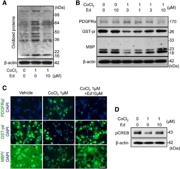
Background and purpose: White matter injury caused by cerebral hypoperfusion may contribute to the pathophysiology of vascular dementia and stroke, but the underlying mechanisms remain to be fully defined. Here, we test the hypothesis that oxidative stress interferes with endogenous white matter repair by disrupting renewal processes mediated by oligodendrocyte precursor cells (OPCs).

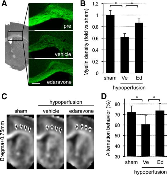
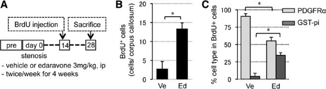
Methods: In vitro, primary rat OPCs were exposed to sublethal CoCl2 for 7 days to induce prolonged chemical hypoxic stress. Then, OPC proliferation/differentiation was assessed. In vivo, prolonged cerebral hypoperfusion was induced by bilateral common carotid artery stenosis in mice. Then, reactive oxygen species production, myelin density, oligodendrocyte versus OPC counts, and cognitive function were evaluated. To block oxidative stress, OPCs and mice were treated with the radical scavenger edaravone.

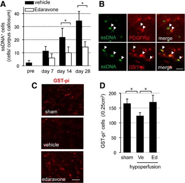
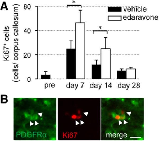
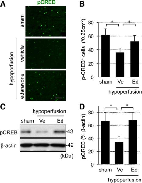
Results: Prolonged chemical hypoxic stress suppressed OPC differentiation in vitro. Radical scavenging with edaravone ameliorated these effects. After 28 days of cerebral hypoperfusion in vivo, reactive oxygen species levels were increased in damaged white matter, along with the suppression of OPC-to-oligodendrocyte differentiation and loss of myelin staining. Concomitantly, mice showed functional deficits in working memory. Radical scavenging with edaravone rescued OPC differentiation, ameliorated myelin loss, and restored working memory function.

Conclusions: Our proof-of-concept study demonstrates that after prolonged cerebral hypoperfusion, oxidative stress interferes with white matter repair by disrupting OPC renewal mechanisms. Radical scavengers may provide a potential therapeutic approach for white matter injury in vascular dementia and stroke.

Keywords: mice; oligodendrocyte; reactive oxygen species; white matter diseases.


Figures

Figure 1
Figure 1
Oxidative stress disturbed oligodendrocyte precursor cell (OPC) differentiation in vitro. A, Representative oxyblot image of 7-day CoCl2-treated OPCs with or without edaravone. B, Representative Western blot images of 7-day CoCl2-treated OPCs with or without edaravone. PDGF-R-α, OPC marker; GST-pi and MBP, oligodendrocyte marker. C, Representative images of PDGF-R-α, GST-pi, and MBP staining in cultured rat OPCs. PDGFRα, GST-pi, MBP: green, DAPI: blue. Scale bar, 25 μm. D, pCREB Western blot image of 7-day CoCl2-treated OPCs with or without edaravone (see Figure II in the online-only Data Supplement for quantitative results of Figure 1A, 1B, and 1D). DAPI indicates 4',6-diamidino-2-phenylindole; Ed, edaravone treatment; GST, glutathione S-transferase; MBP, myelin basic protein; pCREB, phospho-cAMP response element-binding; and PDGF-R-α, platelet-derived growth factor-receptor-α.
Figure 2
Figure 2
Prolonged cerebral hypoperfusion stress induced oxidative stress and white matter dysfunction. A, Representative fluoromyelin staining images of the white matter region (corpus callosum) at day 28. Scale bar, 100 μm. B, Relative intensity of fluoromyelin signals in corpus callosum. C, MR images (bregma +0.75 mm) of sham animals and vehicle- or edaravone-treated white matter injury groups at day 28. D, Alternation behavior (index of working/spatial memory) at day 28 in Y-maze test. Values are mean±SD. n=5 for AC and n=10 for D. Ed indicates edaravone treatment group; and Ve, vehicle. *P<0.05.
Figure 3
Figure 3
Oxidative stress induced cell death in oligodendrocytes and oligodendrocyte precursors in prolonged cerebral hypoperfusion mice. A, Numbers of single-stranded DNA (ssDNA)–positive (ssDNA+) cells in corpus callosum. B, ssDNA staining with a oligodendrocyte precursor cell marker (PDGF-R-α) or a mature oligodendrocyte marker (GST-pi) in the vehicle-treated group at day 28. Scale bar, 10 μm. C, Representative images of GST-pi staining at day 28. Scale bar, 25 μm. D, Numbers of GST-pi–positive (GST-pi+) cells in the lateral side of corpus callosum at day 28. Values are mean±SD of n=5. Ed indicates edaravone treatment group; GST, glutathione S-transferase; PDGF-R-α, platelet-derived growth factor-receptor-α; and Ve, vehicle. *P<0.05.
Figure 4
Figure 4
Reactive oxygen species suppression promoted compensative oligodendrocyte precursor cell proliferation. A, Numbers of Ki67-positive (Ki67+) cells in the white matter. B, Double staining of Ki67 with PDGF-R-α in the edaravone-treated group at day 7. Scale bar, 10 μm. Values are mean±SD of n=5. Ed indicates edaravone treatment group; PDGF-R-α, platelet-derived growth factor-receptor-α; and Ve, vehicle. *P<0.05.
Figure 5
Figure 5
Reactive oxygen species suppression promoted in vivo oligodendrocyte precursor cell differentiation under hypoxic conditions. A, Experimental schedule of cell differentiation assay in vivo. B, Numbers of BrdU-positive (BrdU+) cells in corpus callosum at day 28 (BrdU was incorporated in proliferating cells at day 14). C, Ratio of BrdU/PDGF-R-α or BrdU/GST-pi double-labeled cells in total BrdU+ cells at day 28. Values are mean±SD of n=5. BrdU indicates 5-bromo-2'-deoxyuridine; Ed, edaravone treatment group; GST, glutathione S-transferase; PDGF-R-α, platelet-derived growth factor-receptor-α; and Ve, vehicle. *P<0.05.
Figure 6
Figure 6
Reactive oxygen species suppression promoted CREB activation in white matter brains. A, Representative images of pCREB staining in the white matter at day 28. Scale bar, 25 μm. B, Numbers of pCREB-positive (pCREB+) cells in the lateral side of corpus callosum at day 28. C and D, pCREB Western blot images in the white matter at day 28. β-actin is an internal control. Values are mean±SD of n=5. CREB indicates cAMP response element-binding; Ed, edaravone treatment group; pCREB, phospho-CREB; and Ve, vehicle. *P<0.05.

References

    1. Zhang K, Sejnowski TJ. A universal scaling law between gray matter and white matter of cerebral cortex. Proc Natl Acad Sci U S A. 2000;97:5621–5626. - PMC - PubMed
    1. Ravindranath V, Shivakumar BR, Anandatheerthavarada HK. Low glutathione levels in brain regions of aged rats. Neurosci Lett. 1989;101:187–190. - PubMed
    1. Sasaki T, Senda M. Evaluation of glutathione localization in brain using 99mTc meso-HMPAO. J Nucl Med. 1999;40:1056–1060. - PubMed
    1. Konat GW, Wiggins RC. Effect of reactive oxygen species on myelin membrane proteins. J Neurochem. 1985;45:1113–1118. - PubMed
    1. Smith KJ, Kapoor R, Felts PA. Demyelination: the role of reactive oxygen and nitrogen species. Brain Pathol. 1999;9:69–92. - PMC - PubMed

Publication types

MeSH terms