Review
. 2014 Mar;63(3):185-95.
doi: 10.1097/FJC.0000000000000003.

The immune system and the remodeling infarcted heart: cell biological insights and therapeutic opportunities

Affiliations
Review

The immune system and the remodeling infarcted heart: cell biological insights and therapeutic opportunities

Nikolaos G Frangogiannis. J Cardiovasc Pharmacol. 2014 Mar.

Abstract

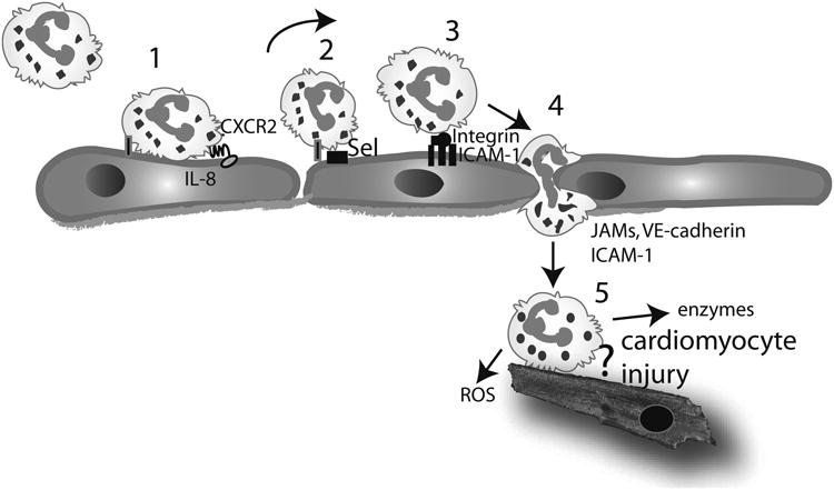
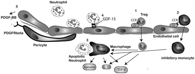
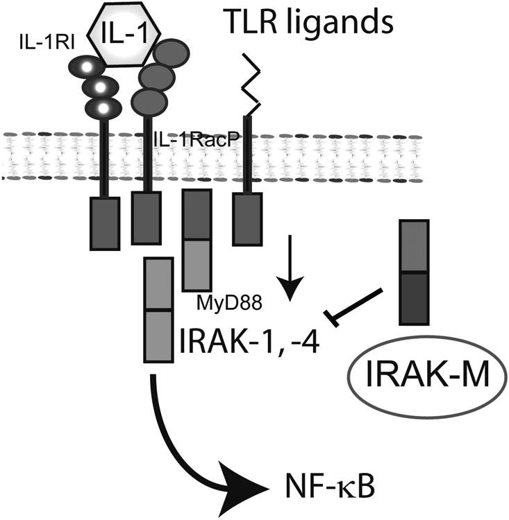
Extensive necrosis of ischemic cardiomyocytes in the infarcted myocardium activates the innate immune response triggering an intense inflammatory reaction. Release of danger signals from dying cells and damaged matrix activates the complement cascade and stimulates Toll-like receptor/interleukin-1 signaling, resulting in the activation of the nuclear factor-κB system and induction of chemokines, cytokines, and adhesion molecules. Subsequent infiltration of the infarct with neutrophils and mononuclear cells serves to clear the wound from dead cells and matrix debris, while stimulating reparative pathways. In addition to its role in repair of the infarcted heart and formation of a scar, the immune system is also involved in adverse remodeling of the infarcted ventricle. Overactive immune responses and defects in suppression, containment, and resolution of the postinfarction inflammatory reaction accentuate dilative remodeling in experimental models and may be associated with chamber dilation, systolic dysfunction, and heart failure in patients surviving a myocardial infarction. Interventions targeting the inflammatory response to attenuate adverse remodeling may hold promise in patients with myocardial infarction that exhibit accentuated, prolonged, or dysregulated immune responses to the acute injury.


Figures

Figure 1
Figure 1
Neutrophil infiltration in the infarcted myocardium and the concept of leukocyte-mediated cardiomyocyte injury. Neutrophil extravasation in the infarcted heart occurs in a series of sequential steps. Circulating neutrophils are captured by activated endothelial cells (1) and roll on the endothelial surface (2) through selectin-mediated interactions. Captured neutrophils can “sense” chemokines immobilized in the endothelial surface (such as IL-8). Interactions between the chemokines and their corresponding chemokine receptors activate integrins on the surface of the neutrophils. Interactions between leukocyte b2 integrins and endothelial adhesion molecules (such as ICAM-1) result in firm adhesion of the neutrophils (3). Subsequently the neutrophils transmigrate across the endothelial layer through interactions that involve adhesion molecules, including VE-cadherin, ICAM-1 and members of the JAM family (4). In the infarcted tissue, neutrophils release reactive oxygen species (ROS) and proteolytic enzymes, playing a role in clearance of the infarct from dead cells and matrix debris (5). Experimental evidence has suggested that neutrophils may adhere to viable cardiomyocytes and exert cytotoxic effects; however, this concept has been challenged by recent studies using genetically targeted animals.
Figure 2
Figure 2
Cellular events associated with negative regulation of the post-infarction inflammatory response. Recruitment of inhibitory mononuclear cell subpopulations in the infarcted myocardium, such as Tregs (1) and inhibitory monocytes (2) may require activation of specific chemokine/chemokine receptor interactions. Macrophages acquire an anti-inflammatory phenotype and secrete inhibitory mediators upon phagocytosis of apoptotic neutrophils (3). Endothelial cells may participate in suppression of the inflammatory response (4) by producing mediators that inhibit adhesive interactions with leukocytes (such as GDF-15). Acquisition of a pericyte coat by infarct neovessels (5) may also suppress inflammatory activity and is mediated through PDGFR-b/PDGF-BB interactions.
Figure 3
Figure 3
Negative regulation of IL-1/TLR responses plays an important role in restraining the post-infarction inflammatory reaction. IRAK-M is expressed in infarct macrophages and fibroblasts and limits IL-1/TLR-mediated NF-κB activation.

References

    1. Frangogiannis NG. The mechanistic basis of infarct healing. Antioxid Redox Signal. 2006 Nov-Dec;8(11-12):1907–1939. - PubMed
    1. Frangogiannis NG. The immune system and cardiac repair. Pharmacol Res. 2008 Aug;58(2):88–111. - PMC - PubMed
    1. Frangogiannis NG. Regulation of the inflammatory response in cardiac repair. Circ Res. 2012 Jan 6;110(1):159–173. - PMC - PubMed
    1. Frantz S, Bauersachs J, Ertl G. Post-infarct remodelling: contribution of wound healing and inflammation. Cardiovasc Res. 2009 Feb 15;81(3):474–481. - PMC - PubMed
    1. Entman ML, Michael L, Rossen RD, Dreyer WJ, Anderson DC, Taylor AA, Smith CW. Inflammation in the course of early myocardial ischemia. Faseb J. 1991 Aug;5(11):2529–2537. - PubMed

Publication types