

Lower peak bone mass and abnormal trabecular and cortical microarchitecture in young men infected with HIV early in life
- PMID: 24072196
- PMCID: PMC4019223
- DOI: 10.1097/QAD.0000000000000070
Lower peak bone mass and abnormal trabecular and cortical microarchitecture in young men infected with HIV early in life
Abstract
Introduction: HIV infection and antiretroviral therapy (ART) early in life may interfere with acquisition of peak bone mass, thereby increasing fracture risk in adulthood.
Methods: We conducted a cross-sectional study of dual-energy X-ray absorptiometry (DXA) and high-resolution peripheral quantitative computed tomography (HR-pQCT) in 30 HIV-infected African-American or Hispanic Tanner stage 5 men aged 20-25 on ART (15 perinatally infected and 15 infected during adolescence) and 15 HIV-uninfected controls.
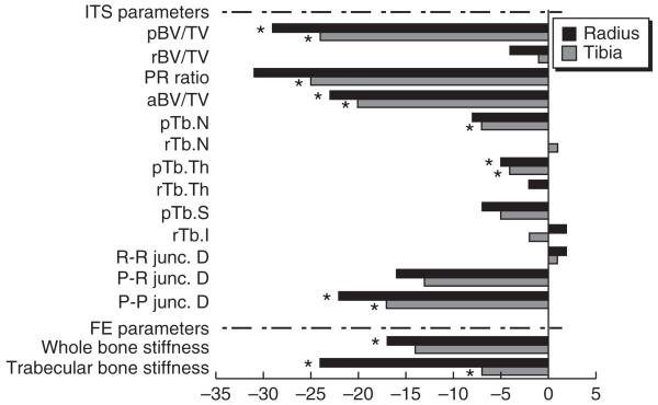
Results: HIV-infected men were similar in age and BMI, but were more likely to be African-American (P = 0.01) than uninfected men. DXA-derived areal bone mineral density (aBMD) Z-scores were 0.4-1.2 lower in HIV-infected men at the spine, hip, and radius (all P < 0.05). At the radius and tibia, total and trabecular volumetric BMD (vBMD), and cortical and trabecular thickness were between 6 and 19% lower in HIV-infected than uninfected men (P <0.05). HIV-infected men had dramatic deficiencies in plate-related parameters by individual trabeculae segmentation (ITS) analyses and 14-17% lower bone stiffness by finite element analysis. Differences in most HR-pQCT parameters remained significant after adjustment for race/ethnicity. No DXA or HR-pQCT parameters differed between men infected perinatally or during adolescence.
Conclusion: At an age by which young men have typically acquired peak bone mass, HIV-infected men on ART have lower BMD, markedly abnormal trabecular plate and cortical microarchitecture, and decreased whole bone stiffness, whether infected perinatally or during adolescence. Reduced bone strength in young adults infected with HIV early in life may place them at higher risk for fractures as they age.
Figures


References
-
- Heaney RP, Abrams S, Dawson-Hughes B, Looker A, Marcus R, Matkovic V, et al. Peak bone mass. Osteoporos Int. 2000;11:985–1009. - PubMed
-
- Jacobson DL, Spiegelman D, Duggan C, Weinberg GA, Bechard L, Furuta L, et al. Predictors of bone mineral density in human immunodeficiency virus-1 infected children. J Pediatr Gastroenterol Nutr. 2005;41:339–346. - PubMed
-
- Arpadi SM, Horlick M, Thornton J, Cuff PA, Wang J, Kotler DP. Bone mineral content is lower in prepubertal HIV-infected children. J Acquir Immune Defic Syndr. 2002;29:450–454. - PubMed
-
- O’Brien KO, Razavi M, Henderson RA, Caballero B, Ellis KJ. Bone mineral content in girls perinatally infected with HIV. Am J Clin Nutr. 2001;73:821–826. - PubMed
Publication types
MeSH terms
Grants and funding
LinkOut - more resources
Full Text Sources
Other Literature Sources
Medical
