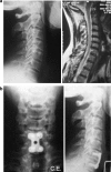
Anterior cervical fusion for radicular-disc conflict performed by three different procedures: clinical and radiographic analysis at long-term follow-up
- PMID: 24072338
- PMCID: PMC3830031
- DOI: 10.1007/s00586-013-3006-z
Anterior cervical fusion for radicular-disc conflict performed by three different procedures: clinical and radiographic analysis at long-term follow-up
Abstract
Purpose: Purpose of the study was to analyze in a retrospective way the clinical and radiographic outcome of three different surgical techniques in patients who underwent anterior cervical fusion.
Methods: Eighty-six patients affected by symptomatic cervical disc herniation or spondylosis underwent cervical anterior fusion. Patients were divided in three groups considering the surgical technique. Clinical outcomes were evaluated by Visual Analog Scale, Odom's criteria, Neck Disability Index. Radiographic evaluation included standard and functional X-rays.
Results: At 7 years mean follow-up, a comparable improvement in clinical symptoms was observed in all groups. Radiographic findings showed a solid fusion in all patients but seven cases in group 2 showed a subsidence of the cage.
Conclusions: As shown by the obtained clinical and radiographic results, the anterior interbody fusion with stand-alone peek cage containing β-tricalcium phosphate could be considered an effective and reliable procedure.
Figures


References
-
- Robinson RA, Smith GW. Anterolateral disc removal and interbody fusion for cervical disc syndrome. Bull Johns Hopkins Hosp. 1955;96:223–224.
-
- Morsher E, Sutter F, Jenny H, et al. Anterior plating of the cervical spine with the hollow screw-plate system of titanium. Chirurg. 1986;57:702–707. - PubMed
-
- Orozco DR, Llovet TR. Osteosintesis en las lesions traumaticas y degeneratives de la columna vertebral. Revista Traumatol Chirurg Rehabil. 1971;1:45–52.
MeSH terms
LinkOut - more resources
Full Text Sources
Other Literature Sources
Medical
Research Materials

