

Comparison of PET imaging with a (68)Ga-labelled PSMA ligand and (18)F-choline-based PET/CT for the diagnosis of recurrent prostate cancer
- PMID: 24072344
- PMCID: PMC3843747
- DOI: 10.1007/s00259-013-2525-5
Comparison of PET imaging with a (68)Ga-labelled PSMA ligand and (18)F-choline-based PET/CT for the diagnosis of recurrent prostate cancer
Abstract
Purpose: Positron emission tomography (PET) with choline tracers has found widespread use for the diagnosis of prostate cancer (PC). However, choline metabolism is not increased in a considerable number of cases, whereas prostate-specific membrane antigen (PSMA) is overexpressed in most PCs. Therefore, a (68)Ga-labelled PSMA ligand could be superior to choline tracers by obtaining a high contrast. The aim of this study was to compare such a novel tracer with standard choline-based PET/CT.
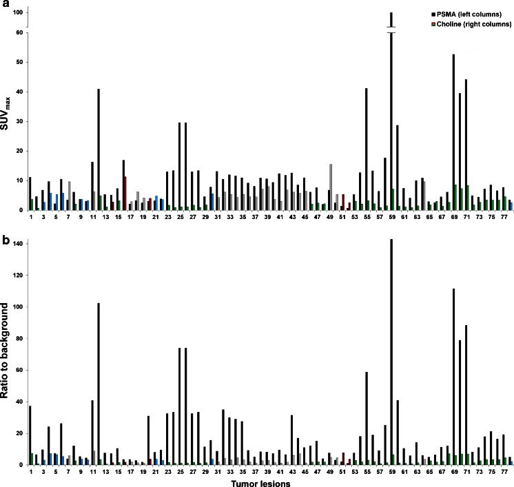
Methods: Thirty-seven patients with biochemical relapse of PC [mean prostate-specific antigen (PSA) 11.1 ± 24.1 ng/ml, range 0.01-116] were retrospectively analysed after (18)F-fluoromethylcholine and (68)Ga-PSMA PET/CT within a time window of 30 days. Radiotracer uptake that was visually considered as PC was semi-quantitatively analysed by measuring the maximum standardized uptake values (SUVmax) of the scans acquired 1 h after injection of (68)Ga-PSMA complex solution (median 132 MBq, range 59-263 MBq) and (18)F-fluoromethylcholine (median 237 MBq, range 114-374 MBq), respectively. In addition, tumour to background ratios were calculated.
Results: A total of 78 lesions characteristic for PC were detected in 32 patients using (68)Ga-PSMA PET/CT and 56 lesions were detected in 26 patients using choline PET/CT. The higher detection rate in (68)Ga-PSMA PET/CT was statistically significant (p=0.04). In five patients no lesion was found with both methods. All lesions detected by (18)F-fluoromethylcholine PET/CT were also seen by (68)Ga-PSMA PET/CT. In (68)Ga-PSMA PET/CT SUVmax was clearly (>10 %) higher in 62 of 78 lesions (79.1 %) and the tumour to background ratio was clearly (>10 %) higher in 74 of 78 lesions (94.9 %) when compared to (18)F-fluoromethylcholine PET/CT.
Conclusion: (68)Ga-PSMA PET/CT can detect lesions characteristic for PC with improved contrast when compared to standard (18)F-fluoromethylcholine PET/CT, especially at low PSA levels.
Figures
Comment in
-
Writing PET into existence.Eur J Nucl Med Mol Imaging. 2014 Jan;41(1):7-10. doi: 10.1007/s00259-013-2573-x. Epub 2013 Sep 27. Eur J Nucl Med Mol Imaging. 2014. PMID: 24072349 No abstract available.
References
-
- Kosuri S, Akhtar NH, Smith M, Osborne JR, Tagawa ST. Review of salvage therapy for biochemically recurrent prostate cancer: the role of imaging and rationale for systemic salvage targeted anti-prostate-specific membrane antigen radioimmunotherapy. Adv Urol. 2012;2012:921674. doi: 10.1155/2012/921674. - DOI - PMC - PubMed
-
- Igerc I, Kohlfürst S, Gallowitsch HJ, Matschnig S, Kresnik E, Gomez-Segovia I, et al. The value of 18F-choline PET/CT in patients with elevated PSA-level and negative prostate needle biopsy for localisation of prostate cancer. Eur J Nucl Med Mol Imaging. 2008;35:976–983. doi: 10.1007/s00259-007-0686-9. - DOI - PubMed
Publication types
MeSH terms
Substances
LinkOut - more resources
Full Text Sources
Other Literature Sources
Medical
Research Materials
Miscellaneous
