Review
. 2013 Nov;15(11):375.
doi: 10.1007/s11926-013-0375-6.

Homeostatic mechanisms in articular cartilage and role of inflammation in osteoarthritis

Affiliations
Review

Homeostatic mechanisms in articular cartilage and role of inflammation in osteoarthritis

Xavier Houard et al. Curr Rheumatol Rep. 2013 Nov.

Abstract

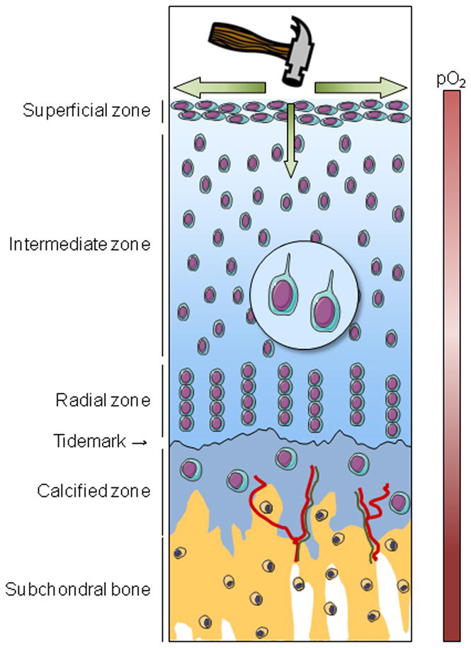
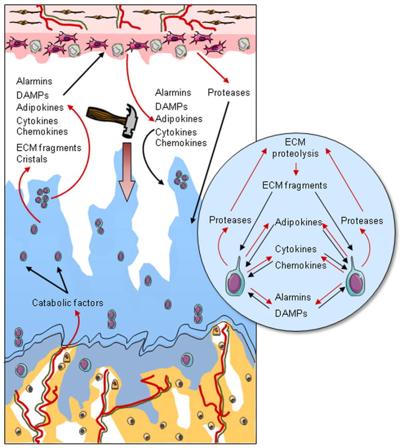
Osteoarthritis (OA) is a whole joint disease, in which thinning and disappearance of cartilage is a critical determinant in OA progression. The rupture of cartilage homeostasis whatever its cause (aging, genetic predisposition, trauma or metabolic disorder) induces profound phenotypic modifications of chondrocytes, which then promote the synthesis of a subset of factors that induce cartilage damage and target other joint tissues. Interestingly, among these factors are numerous components of the inflammatory pathways. Chondrocytes produce cytokines, chemokines, alarmins, prostanoids, and adipokines and express numerous cell surface receptors for cytokines and chemokines, as well as Toll-like receptors. These receptors activate intracellular signaling pathways involved in inflammatory and stress responses of chondrocytes in OA joints. This review focuses on mechanisms responsible for the maintenance of cartilage homeostasis and highlights the role of inflammatory processes in OA progression.


Figures

Figure 1
Figure 1. Schematic representation of cartilage organization in healthy joint
Healthy articular cartilage comprises four different areas: the superficial, intermediate, radial and calcified zones. Each is characterized by a peculiar chondrocyte phenotype and by distinctive extracellular matrix organization and composition. The calcified zone differs from the three other zones by the mineralization of its extracellular matrix, by the presence of vessels (red) and by nerve fibers (green) that originate from the subchondral bone. The calcified zone interfaces with the non-mineralized cartilage, from which it is separated by the tidemark, and the subchondral bone. Due to the absence of vessels within cartilage, chondrocytes live in a hypoxic environment. Hypoxia is important for chondrocyte function and viability. Oxygen and nutrients come from the vascular supply in the synovium and the subchondral bone. A main function of cartilage is the absorption and the dissipation of mechanical load, which is necessary to maintain cartilage homeostasis. The primary cilium plays a crucial role in cartilage homeostasis, especially in the perception of mechanical load due to the presence of integrins and ion channels.
Figure 2
Figure 2. Schematic representation of cartilage alterations in OA
In OA, there is a progressive disappearance of cartilage associated with chondrocyte loss and phenotypic modifications, including the formation of clusters, the activation of a catabolic phenotypic and hypertrophic differentiation. In addition to cartilage damage, remodeling of the subchondral bone occurs with the development of vessels (red) located in structures called vascular channels, which also contain osteoblasts, osteoclasts, and sensory nerves (green). Vascular channels are supposed to facilitate biochemical communication between the bone and the cartilage. In response to several stimuli, including inappropriate mechanical loading and catabolic factors coming from the subchondral bone, chondrocytes modify their phenotype and express a subset of factors, such as cytokines, chemokines, alarmins, DAMPs, and adipokines. All of these mediators act as paracrine factors and initiate a vicious circle of cartilage degradation but also reach the synovium and provoke an inflammatory process with the production by synovial macrophages and fibroblasts of factors, which both promote inflammation in the synovium and participate in cartilage damage.

References

    1. Blagojevic M, Jinks C, Jeffery A, Jordan KP. Risk factors for onset of osteoarthritis of the knee in older adults: a systematic review and meta-analysis. Osteoarthritis Cartilage. 2010;18(1):24–33. - PubMed
    1. Felson DT, Lawrence RC, Dieppe PA, et al. Osteoarthritis: new insights Part 1: the disease and its risk factors. Ann Intern Med. 2010;133(8):635–646. - PubMed
    1. Mahjoub M, Berenbaum F, Houard X. Why subchondral bone in osteoarthritis? The importance of the cartilage bone interface in osteoarthritis. Osteoporos Int. 2012;23(Suppl 8):841–846. - PubMed
    1. Lyons TJ, McClure SF, Stoddart RW, McClure J. The normal human chondro-osseous junctional region: evidence for contact of uncalcified cartilage with subchondral bone and marrow spaces. BMC Musculoskelet Disord. 2006;7:52. - PMC - PubMed
    1. Fawns HT, Landells JW. Histochemical studies of rheumatic conditions. I. Observations on the fine structures of the matrix of normal bone and cartilage. Ann Rheum Dis. 1953;12(2):105–113. - PMC - PubMed

Publication types

MeSH terms