. 2013 Nov 8;288(45):32809-32820.
doi: 10.1074/jbc.M113.512962. Epub 2013 Sep 26.

Stabilization of speckle-type POZ protein (Spop) by Daz interacting protein 1 (Dzip1) is essential for Gli turnover and the proper output of Hedgehog signaling

Affiliations

Stabilization of speckle-type POZ protein (Spop) by Daz interacting protein 1 (Dzip1) is essential for Gli turnover and the proper output of Hedgehog signaling

Tyler Schwend et al. J Biol Chem. .

Abstract

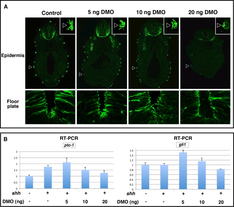
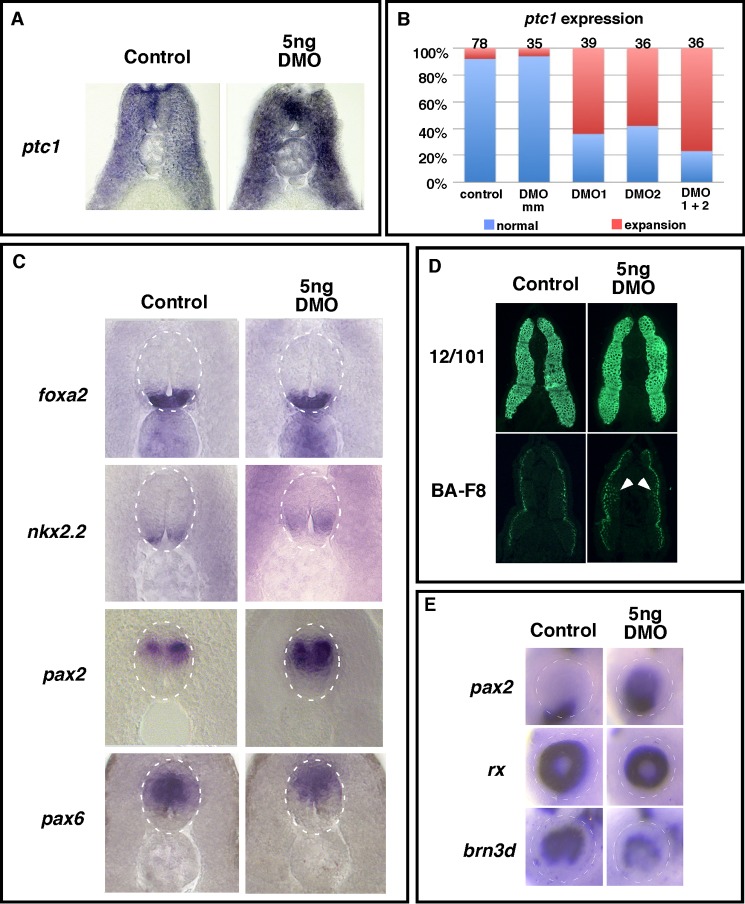
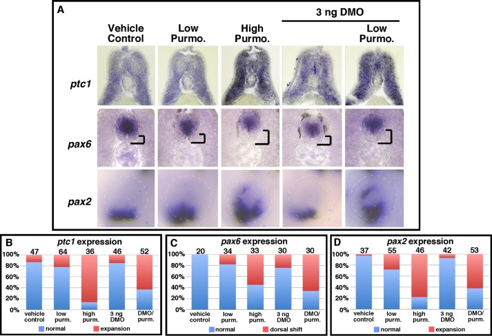
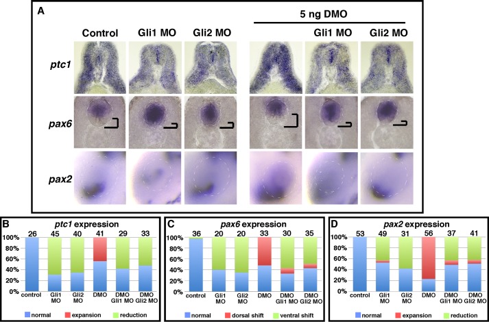
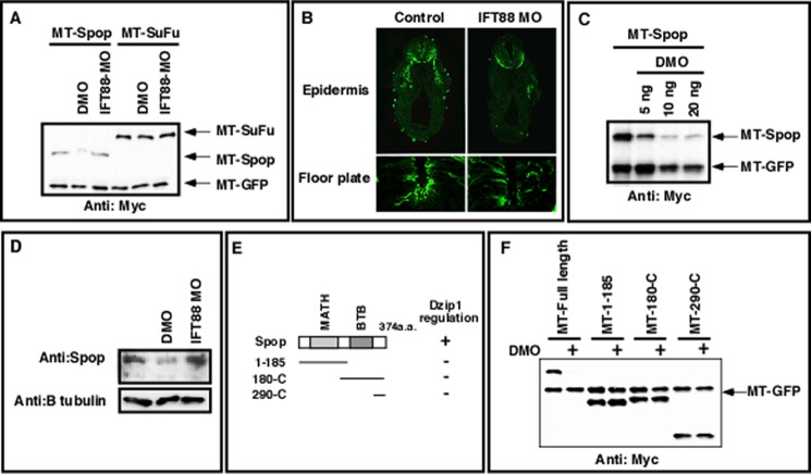
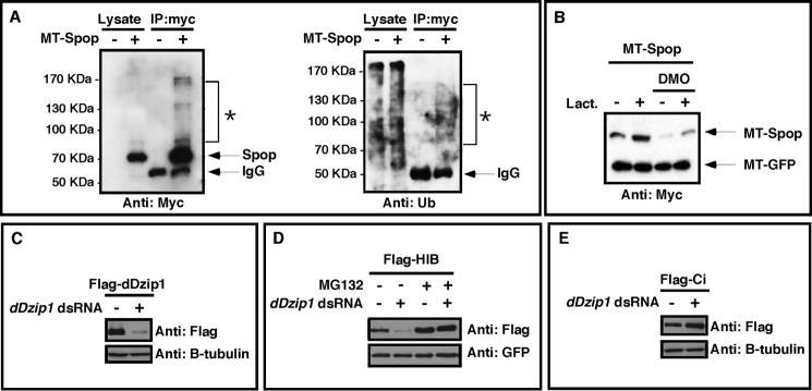
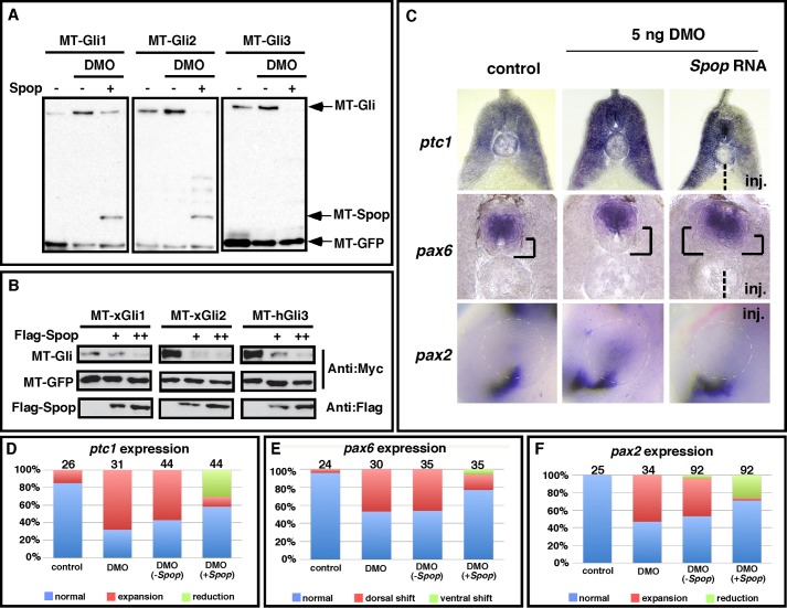
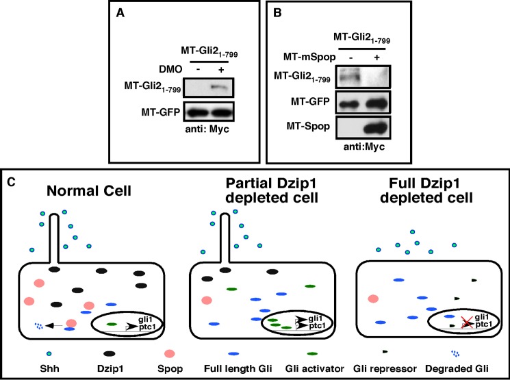
The Hedgehog (Hh) pathway is essential for embryonic development and adult tissue homeostasis. The Gli/Cubitus interruptus (Ci) family of transcription factors acts at the downstream end of the pathway to mediate Hh signaling. Both Hh-dependent and -independent Gli regulatory mechanisms are important for the output of Hh signaling. Daz interacting protein 1 (Dzip1) has bipartite positive and negative functions in the Hh pathway. The positive Hh regulatory function appears to be attributed to a requirement for Dzip1 during ciliogenesis. The mechanism by which Dzip1 inhibits Hh signaling, however, remains largely unclear. We recently found that Dzip1 is required for Gli turnover, which may account for its inhibitory function in Hh signaling. Here, we report that Dzip1 regulates Gli/Ci turnover by preventing degradation of speckle-type POZ protein (Spop), a protein that promotes proteasome-dependent turnover of Gli proteins. We provide evidence that Dzip1 regulates the stability of Spop independent of its function in ciliogenesis. Partial knockdown of Dzip1 to levels insufficient for perturbing ciliogenesis, sensitized Xenopus embryos to Hh signaling, leading to phenotypes that resemble activation of Hh signaling. Importantly, overexpression of Spop was able to restore proper Gli protein turnover and rescue phenotypes in Dzip1-depleted embryos. Consistently, depletion of Dzip1 in Drosophila S2 cells destabilized Hh-induced BTB protein (HIB), the Drosophila homolog of Spop, and increased the level of Ci. Thus, Dzip1-dependent stabilization of Spop/HIB is evolutionarily conserved and essential for proper regulation of Gli/Ci proteins in the Hh pathway.

Keywords: Cilia; Development; Dzip1; Gli; Hedgehog; Protein Degradation; Spop; Xenopus.


Figures

FIGURE 1.
FIGURE 1.
Sensitization of Xenopus animal caps to Shh by partial knockdown of Dzip1. A, immunofluorescence showing primary cilia in the floor plate (bottom panel) and epidermis motile cilia (top panel, arrowheads) in control (left column) and embryos injected with 5, 10, or 20 ng of DMOs (from left to right). B, real-time RT-PCR showing the expression of ptc1 and gli1 in animal caps from controls and embryos injected with shh mRNA (500pg) alone or in combination with 5, 10, or 20 ng of DMOs. All samples were normalized to odc levels.
FIGURE 2.
FIGURE 2.
Partial depletion of Dzip1 increased the expression of Hh target genes in embryos. A, in situ hybridization showing that partial Dzip1 depletion (right) markedly increased the expression of ptc1 in the neural tube and somites at stage 35. B, summary of the ptc1 expression phenotypes in control and morpholino-injected embryos. DMO mm, 5-bp mismatch control morpholino of DMO2. C, in situ hybridization showing the expression of floor plate markers (foxa2 and nkx2.2) and markers for more dorsal neural tube cells (pax2 and pax6) in controls (left) and embryos injected with DMO (right). The white dotted lines outline the neural tube borders. D, immunofluorescence showing total muscle (12/101 reactivity; top panel) and slow muscle cells (BA-F8 reactivity; bottom panel) in controls (left column) and DMO-injected embryos (right column). Arrowheads denote ectopic BA-F8 reactive slow muscle cells evident in DMO injected embryos. E, in situ hybridization showing eye patterning in controls (left) and DMO injected (right) embryos. Markers for optic stalk (pax2, top panel), total retina (rx, middle panel), and dorsal retina (brn3d, bottom panel) were analyzed. Dotted lines outline the eye.
FIGURE 3.
FIGURE 3.
Partial depletion of Dzip1 sensitizes embryos to the Smo agonist purmorphamine. A, whole mount in situ hybridization of ptc1 (top), pax6 (middle), and pax2 (bottom) in vehicle-treated (ethanol) controls and embryos treated with either a low dose of purmorphamine (2.5–5.0 μm), a high dose of purmorphamine (10 μm), injected with a low concentration of DMO (3 ng), or receiving a combination of a low dose of purmorphamine (Purmo. or purm.) and an injection with a low concentration of DMO. In the middle panels, the distance between the floor plate and the ventral border of the pax6 expression domain was highlighted. B–D, summary of ptc1 expression phenotypes (B), pax6 expression phenotypes (C), or pax2 expression phenotypes (D) for vehicle-treated (ethanol) controls and the different groups of embryos subjected to purmorphamine, DMO, or both.
FIGURE 4.
FIGURE 4.
Knockdown of Gli1 or Gli2 rescues phenotypes induced by partial depletion of Dzip1. A, whole mount in situ hybridization of ptc1 (top), pax6 (middle), and pax2 (bottom) in controls and embryos receiving either a single injection of Gli1 MO, Gli2 MO, or DMO and double knockdown embryos injected with Gli1 MO/DMO or Gli2 MO/DMO. In the middle panels, the distance between the floor plate and the ventral border of the pax6 expression domain was highlighted. B--D, summary of ptc1 expression phenotypes (B), pax6 expression phenotypes (C), or pax2 expression phenotypes (D) in controls and the different groups of embryos receiving either a single or double morpholino injection against Gli1, Gli2, or Dzip1.
FIGURE 5.
FIGURE 5.
Dzip1 regulates the stability of Spop independent of its role in ciliogenesis. A, Western blot showing that the stability of Spop, but not that of Sufu, was decreased in Dzip1-depleted embryos. RNAs encoding MT-Spop (250 pg) or MT-Sufu (250 pg) were co-injected with MT-GFP (50 pg) into controls or embryos that had received a prior injection of DMO or IFT88 morpholino (MO). Embryos were harvested at the neurula stage. MT-GFP serves as injection and loading control. B, immunofluorescence showing a significant reduction of motile cilia on the epidermis (top panel) and primary cilia in the floor plate (bottom panel) within embryos injected with 40 ng IFT88 morpholino (right column) compared with controls (left column). C, DMO injection negatively impacted Spop stability in a dose-dependent fashion. MT-Spop was injected with MT-GFP into controls or embryos that had received increasing concentrations of DMO (5–20 ng). Expression of Spop was assessed by Western blotting. D, endogenous Spop protein was reduced in Dzip1-depleted embryos and unchanged in IFT88-depleted embryos. Embryos were harvested at stage 18 and subjected to Western blot with the anti-Spop antibody to detect endogenous Spop levels. Detection of β-tubulin levels serves as a loading control. E, schematic presentation of Spop deletion constructs. All deletion constructs were N-terminally myc-tagged. F, Western blot results showing the effects of Dzip1 knockdown on the stability of full-length and deletions of Spop. All constructs were co-injected with MT-GFP. Embryo lysates were subjected to Western blot with the anti-myc antibody.
FIGURE 6.
FIGURE 6.
Dzip1 prevents ubiquitin/proteasome-dependent degradation of Spop/HIB. A, Spop is ubiquitinated in vivo. MT-Spop was injected into Xenopus embryos. Spop protein was purified by immunoprecipitation using an anti-myc antibody. Lysates and immunoprecipitates (IP) from injected and uninjected embryos were subjected to Western blot with the anti-myc antibody (left panel) and anti-ubiquitin antibody (right panel). The unmodified MT-Spop band migrated to 68 kDa. A smear of high molecular weight proteins (*) migrating slower than unmodified MT-Spop could be detected in both myc and ubiquitin Westerns. B, MT-Spop was injected into control or embryos that had received a prior injection of DMO. Animal caps were dissected at stage 9. Subsequently, caps were cultured in media with or without 2 μg of lactacystin (Lact.). Animal caps were harvested at stage 15 and subjected to Western blot to detect the levels of Spop. C, Western blot results showing severe reduction of FLAG-dDzip1 by dDzip1 dsRNA in S2 cells. β-Tubulin serves as a loading control. D, Dzip1 knockdown promoted proteasome-dependent degradation of HIB in S2 cells. S2 cells were cotransfected with FLAG-HIB and GFP constructs and treated with dDzip1 dsRNA and/or MG132. Cell extracts were subjected to Western blot with the anti-FLAG antibody to detect the levels of HIB. GFP serves as the transfection and loading control. E, Dzip1 knockdown promotes Ci stability in S2 cells. S2 cells were transfected with FLAG-Ci and treated with dDzip1 dsRNA. Western blots were carried out with the cell lysates to determine the levels of Ci. Tubulin serves as a loading control.
FIGURE 7.
FIGURE 7.
Overexpression of Spop rescues phenotypes caused by partial knockdown of Dzip1. A, Western blot results showing that increases to Gli stability after Dzip1 depletion were reversed by Spop overexpression. RNAs encoding MT-Gli1–3 (2 ng) and MT-GFP (50 pg) were injected alone or in combination with MT-Spop (100 pg) into controls or embryos that had received a prior injection of DMO. Embryo were harvested at the neurula stage and subjected to Western blot with the anti-myc antibody to detect the levels of Spop. B, forced Spop expression decreases the stability of xGli1, xGli2, and hGli3 in NIH3T3 cells. NIH3T3 cells were transfected with MT-Glis and MT-GFP alone or in combination with increasing levels of FLAG-Spop. Western blots were carried out with the cell lysates to determine the levels of Gli1, -2, and -3. GFP serves as a transfection and loading control. C, in situ hybridization results showing the expression of ptc1, pax6, and pax2 in controls or embryos injected with DMO alone or DMO and Spop RNA. inj. indicates the side that received Spop RNA injection. D–F, summary of the ptc1 (D), pax6 (E), and pax2 (F) expression phenotypes in controls and injected embryos.
FIGURE 8.
FIGURE 8.
Regulation of Gli21–799, an analog Gli2 mutant of zebrafish Yot, by Dzip1 and Spop. A, Western blot results showing that knockdown of Dzip1 stabilized Gli21–799 in Xenopus embryos. RNAs encoding Gli21–799 and MT-GFP were injected into controls and embryos that had received a prior injection of DMO. Embryos were harvested at the neurula stage and subjected to Western blotting. MT-GFP served as an injection and loading control. B, forced Spop expression decreased the protein stability of Gli21–799. MT-Gli21–799 and MT-GFP were transfected alone or in combination with MT-Spop into NIH3T3 cells. Cell lysates were subjected to Western blot with the anti-myc antibody to detect the levels of Spop. C, model of Dzip1 functions during Hh signaling. Dzip1 supports ciliogenesis and maintains the proper expression level of Gli proteins through controlling the stability of Spop (left panel). Partial knockdown of Dzip1 has no effect on the formation of cilia but induces degradation of Spop, leading to increased levels of Gli proteins. When exposed to Shh, these cells produce more Gli activators, resulting in a more robust Hh target gene expression (middle panel). When Dzip1 was depleted more completely, ciliogenesis was disrupted. Although these cells have increased levels of Gli, they cannot receive Shh. This prevents the formation of Gli activators. Consequently, gene expression induced by Shh were inhibited (right panel). Under normal conditions, cells located at a distance from the source of Shh are not exposed to Shh. Whether these cells form cilia or not are not important. However, when Dizp1 is depleted, destabilization of Spop will induce accumulation of full-length Gli proteins (not Gli activators). This will lead to weak ectopic Hh target gene expression in embryos.

References

    1. Dellovade T., Romer J. T., Curran T., Rubin L. L. (2006) The hedgehog pathway and neurological disorders. Annu. Rev. Neurosci. 29, 539–563 - PubMed
    1. Ingham P. W., McMahon A. P. (2001) Hedgehog signaling in animal development. Paradigms and principles. Genes Dev. 15, 3059–3087 - PubMed
    1. Mullor J. L., Sánchez P., Ruiz i Altaba A. (2002) Pathways and consequences. Hedgehog signaling in human disease. Trends Cell Biol. 12, 562–569 - PubMed
    1. Kasper M., Regl G., Frischauf A. M., Aberger F. (2006) GLI transcription factors. Mediators of oncogenic Hedgehog signalling. Eur. J. Cancer 42, 437–445 - PubMed
    1. Taipale J., Beachy P. A. (2001) The Hedgehog and Wnt signalling pathways in cancer. Nature 411, 349–354 - PubMed

Publication types

MeSH terms

Substances