. 2013 Nov 15;288(46):33398-410.
doi: 10.1074/jbc.M113.499848. Epub 2013 Sep 26.

Characterization of three full-length human nonmuscle myosin II paralogs

Affiliations

Characterization of three full-length human nonmuscle myosin II paralogs

Neil Billington et al. J Biol Chem. .

Abstract

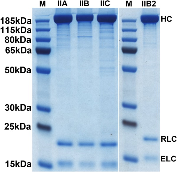
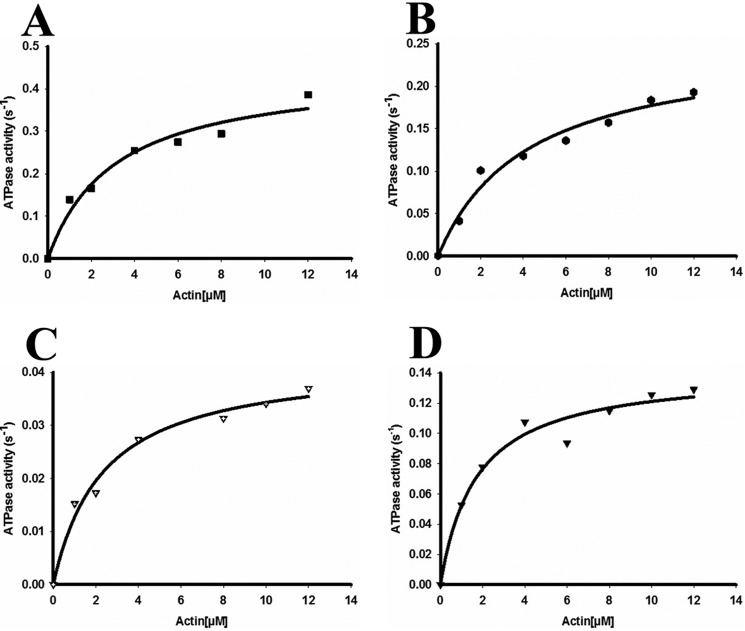
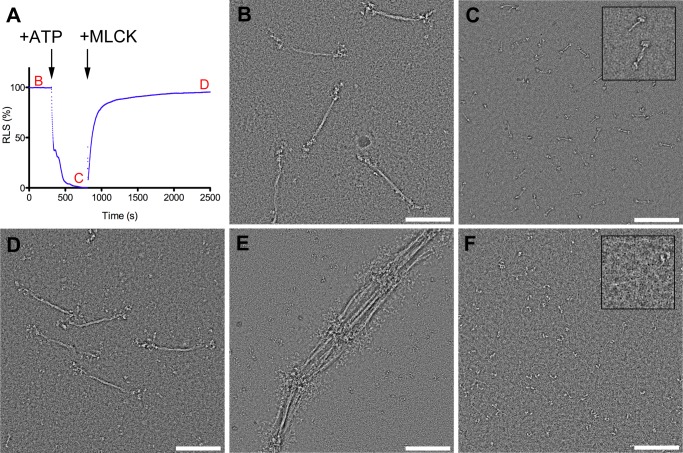
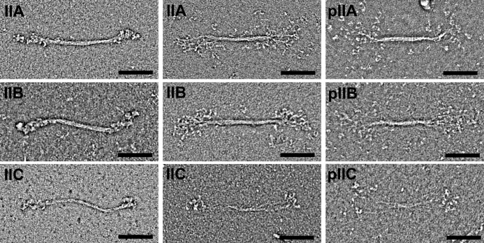
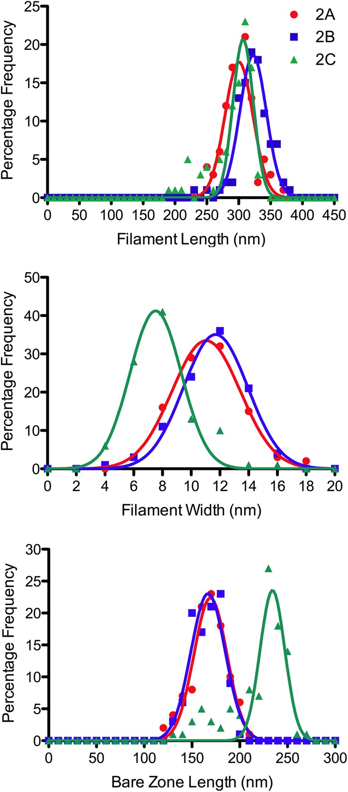
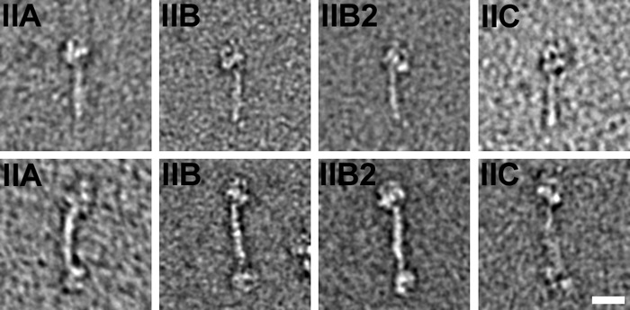
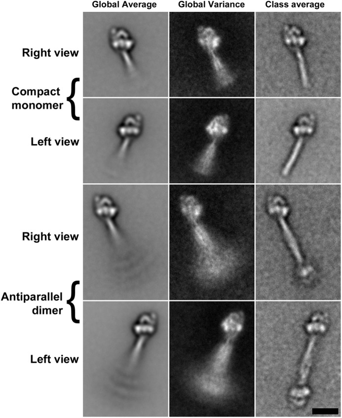
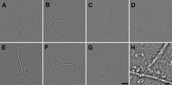
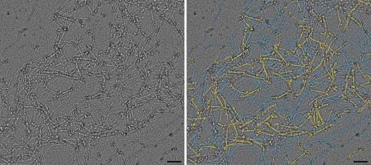
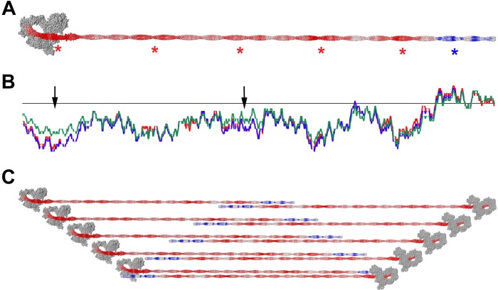
Nonmuscle myosin IIs (NM IIs) are a group of molecular motors involved in a wide variety of cellular processes including cytokinesis, migration, and control of cell morphology. There are three paralogs of the NM II heavy chain in humans (IIA, IIB, and IIC), each encoded by a separate gene. These paralogs are expressed at different levels according to cell type and have different roles and intracellular distributions in vivo. Most previous studies on NM II used tissue-purified protein or expressed fragments of the molecule, which presents potential drawbacks for characterizing individual paralogs of the intact protein in vitro. To circumvent current limitations and approach their native properties, we have successfully expressed and purified the three full-length human NM II proteins with their light chains, using the baculovirus/Sf9 system. The enzymatic and structural properties of the three paralogs were characterized. Although each NM II is capable of forming bipolar filaments, those formed by IIC tend to contain fewer constituent molecules than those of IIA and IIB. All paralogs adopt the compact conformation in the presence of ATP. Phosphorylation of the regulatory light chain leads to assembly into filaments, which bind to actin in the presence of ATP. The nature of interactions with actin filaments is shown with different paralogs exhibiting different actin binding behaviors under equivalent conditions. The data show that although NM IIA and IIB form filaments with similar properties, NM IIC forms filaments that are less well suited to roles such as tension maintenance within the cell.

Keywords: ATPases; Actin; Cytoskeleton; Electron Microscopy (EM); Molecular Motors; Myosin.


Figures

FIGURE 1.
FIGURE 1.
Expression and purification of FLAG-tagged full-length NM IIs from baculovirus-infected Sf9 cells. NM II proteins were purified by FLAG affinity chromatography (see “Experimental Procedures”). Purified NM II heavy chains and two light chains were separated on a 10% (Bis-Tris) polyacrylamide-SDS gel, followed by PageBlue staining. The positions of the NM II heavy chain (HC), RLC, and ELC are shown on the right. M, molecular mass marker (PageRulerTM Plus Prestained).
FIGURE 2.
FIGURE 2.
Enzymatic activity of full-length wild-type NM II molecules. Actin-activated MgATPase activity of full-length wild-type NM IIA (A), NM IIB (B), NM IIB2 (C), and NM IIC (D) was measured at 150 mm KCl at 25 °C (details under “Experimental Procedures”). NM II was phosphorylated with MLCK at room temperature for 30 min prior to the assay. The actin concentration varied from 0 to 12 μm. MgATPase activity of NM II at 0 μm actin concentration was subtracted from each data point (< 0.01 s−1 for all paralogs). Data sets were fitted to a hyperbolic equation to determine the kinetic constants, namely, Vmax and KATPase. The data shown were from the measurement of a single preparation of NM II. Note the different y axis scales.
FIGURE 3.
FIGURE 3.
General assembly behavior of NM II as exemplified by NM IIB. A, time course of relative light scattering (RLS). The arrows indicate the times where ATP and MLCK (with Ca2+ and CaM) were added. Red letters refer to EM images in subsequent panels. B, bipolar thick filaments formed at approximately physiological ionic strength (150 mm KCl) in the absence of ATP. C, addition of 1 mm ATP to a suspension of filaments at physiological ionic strength leads to disassembly into compact individual molecules. Inset shows examples of a compact monomer and antiparallel dimer. D, compact molecules reassemble into filaments following RLC phosphorylation with MLCK. E, lowering the ionic strength to 100 mm KCl leads to lateral and serial aggregation of filaments by their head containing ends. F, in high ionic strength buffer (0.5 m KCl), myosin is extended and unpolymerized. Inset highlights one such molecule.
FIGURE 4.
FIGURE 4.
Appearance of wild-type NM II filaments. The left two columns show unphosphorylated filaments in the absence of nucleotide. The right column shows filaments in which the RLCs are phosphorylated. In the absence of nucleotide, NM IIA, B, and C form filaments with similar gross morphology. In all cases the filaments are bipolar and ∼300 nm in size. Some filaments have a compact appearance where the heads are packed closely against the backbone (left panels), whereas in others a bouquet of heads is seen with individual molecules splayed out from the filament backbone (right panels). The appearances of unphosphorylated and phosphorylated filaments are similar. Scale bar, 100 nm.
FIGURE 5.
FIGURE 5.
Comparison of filament dimensions for NM IIA, IIB, and IIC. Length distributions of filaments from NM II paralogs are similar. Mean contour lengths were 301 (±24 nm S.D.), 323 (±24 nm S.D.), and 293 (±33 nm S.D.) for NM IIA, NM IIB, and NM IIC respectively. NM IIC filaments tended to be narrower and have a longer bare zone than those made from NM IIA and NM IIB, indicating fewer constituent molecules in NM IIC filaments. Mean widths were 11.2 (±2.4 nm S.D.), 11.5 (±2.3 nm), and 7.9 nm (±2.1 nm) for NM IIA, NM IIB, and NM IIC, respectively. Mean bare zone lengths were 167 (±19 nm) for NM IIA, 166 (±16 nm) for NM IIB, and 219 (±30 nm) for NM IIC.
FIGURE 6.
FIGURE 6.
Effect of ATP on unphosphorylated RLC NM II. All three paralogs of NM II, as well as the IIB2 splice variant, form a compact structure following addition of 1 mm ATP (upper panels). In all three paralogs, pairs of compact molecules are associated in an antiparallel dogbone structure (lower panels). Scale bar, 20 nm (applies to all panels).
FIGURE 7.
FIGURE 7.
Image processing reveals the compact NM II structures in more detail. Alignment and classification of images of NM IIB in the presence of 1 mm ATP shows the structure to be very similar to that previously detailed for smooth muscle myosin in which the heads interact in an asymmetric fashion and the tail is folded and wrapped around the heads to form a compact molecule ∼50 nm in length (upper two rows) (44, 53, 54). The dogbone structure is an antiparallel pair of compact molecules in which the folded tail regions of two myosins are associated with a mean overlap of 28 nm (lower two rows). The left column shows global average images, the central column shows global variance images, and the right column shows a selected class average in each case. Scale bar, 20 nm (applies to all panels).
FIGURE 8.
FIGURE 8.
Interactions of phosphorylated RLC NM II with actin in the presence of 1 mm ATP. Following phosphorylation of the RLC with MLCK, NM II from all paralogs (as well as the IIB2 splice variant) forms filaments that are capable of binding to actin. A, NM IIA; B, NM IIB; C, NM IIB2; D, NM IIC. Multiple modes of interaction with actin can be seen. E and F, in addition to the single sided attachments seen in A–D, single myosin filaments can bind multiple actin filaments via a single filament end (E) or via both filament ends (F). G, NM II filaments are also capable of binding to a single actin filament via both ends, although this is less common than the single sided attachment. H, because individual myosins splay away from the filament backbone, a large area of actin, spanning multiple helical repeats, is accessible to the bouquet of heads. E–H show NM IIB2. Scale bar in G, 50 nm (applies to A–G); scale bar in H, 10 nm.
FIGURE 9.
FIGURE 9.
Superprecipitation of actin caused by phosphorylated RLC NM IIB filaments in the presence of ATP. The left panel shows an example of a dense actomyosin network resulting from interaction of 500 nm actin with 50 nm NM IIB. The right panel shows a false color version of the same image highlighting myosin filaments (yellow, myosin; blue, actin).
FIGURE 10.
FIGURE 10.
Model of antiparallel overlaps in NM II filaments. Charge distribution of exposed residues is shown on a model of NM IIB. Charges are smoothed over a surrounding window of 98 residues. Red is negative charge, and blue is positive charge. Asterisks show the positions of strongly charged regions (A). Charge distribution of each NM II paralog along the coiled coil. Red, blue, and green represent charges of NM IIA, NM IIB, and NM IIC, respectively (B). Schematic diagram showing antiparallel overlaps, which allow the positively charged region to interact with the more negatively charged regions of the coiled coil (C). The heads in this figure are displayed in the compact, folded position for visual simplicity rather than to imply compact head packing within the filament.

References

    1. Odronitz F., Kollmar M. (2007) Drawing the tree of eukaryotic life based on the analysis of 2,269 manually annotated myosins from 328 species. Genome Biol. 8, R196. - PMC - PubMed
    1. Richards T. A., Cavalier-Smith T. (2005) Myosin domain evolution and the primary divergence of eukaryotes. Nature 436, 1113–1118 - PubMed
    1. Kühne W. (1864) Untersuchungen über das protplasma und di contractilitat, W. Engelmann, Leipzig, Germany
    1. Craig R., Megerman J. (1977) Assembly of smooth muscle myosin into side-polar filaments. J. Cell Biol. 75, 990–996 - PMC - PubMed
    1. Lo C. M., Buxton D. B., Chua G. C., Dembo M., Adelstein R. S., Wang Y. L. (2004) Nonmuscle myosin IIb is involved in the guidance of fibroblast migration. Mol. Biol. Cell 15, 982–989 - PMC - PubMed

Publication types

MeSH terms