. 2014 Jan;28(1):56-66.
doi: 10.1096/fj.13-231720. Epub 2013 Sep 26.

Inhibition of protein translation as a novel mechanism for prostaglandin E2 regulation of cell functions

Affiliations

Inhibition of protein translation as a novel mechanism for prostaglandin E2 regulation of cell functions

Katsuhide Okunishi et al. FASEB J. 2014 Jan.

Abstract

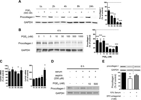
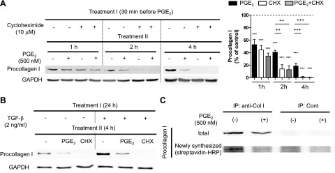
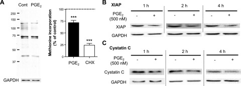
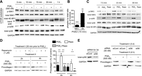
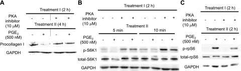
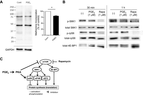
Prostaglandin E2 (PGE2) regulates numerous biological processes by modulating transcriptional activation, epigenetic control, proteolysis, and secretion of various proteins. Scar formation depends on fibroblast elaboration of matrix proteins such as collagen, and this process is strongly suppressed by PGE2 through activation of cAMP-dependent protein kinase A (PKA). However, the actual mechanism by which PGE2-PKA signaling inhibits collagen expression in fibroblasts has never been delineated, and that was the objective of this study. PGE2 unexpectedly induced a rapid reduction in procollagen I protein expression in adult lung fibroblasts, with a half-maximum effect at 1.5 h. This effect reflected its inhibition of translation rather than transcription. Global protein synthesis was also inhibited by PGE2. This action was mediated by PKA and involved both activation of ribosomal protein (rpS6) and suppression of mammalian target of rapamycin (mTOR). Similar effects of PGE2 were demonstrated in mouse peritoneal macrophages (PMs). These findings identify inhibition of translation as a new mechanism by which PGE2 regulates cellular function and a novel example of translational inhibition mediated by opposing actions on two distinct translational control pathways. Translational inhibition would be expected to contribute to dynamic alterations in cell function that accompany the changing PGE2 levels observed in disease states and with various pharmacotherapies.

Keywords: fibroblasts; macrophages; mammalian target of rapamycin; protein kinase A; ribosomal protein S6.


Figures

Figure 1.
Figure 1.
Rapid suppression by PGE2 of procollagen I protein expression in normal adult lung fibroblasts. A–C) After overnight serum starvation, cell culture medium was changed to fresh serum-free medium, and normal adult lung fibroblasts were then incubated with or without PGE2 (at 500 nM in A and at various concentrations in B). A, B) Time course (A) and dose dependency (B) of the effect of PGE2 on procollagen I protein expression. At each time point after addition of PGE2 at the indicated concentrations, cell lysates were harvested, and procollagen I levels in the lysates were determined by immunoblot analysis with densitometry. Left panel: representative immunoblot. Right panel: results of densitometric analysis of procollagen I levels from 3–5 experiments (A) or 3 experiments (B). After normalization to GAPDH, the procollagen I level in PGE2-treated cells was expressed relative to that in cells without PGE2 (expressed as 100%, dashed line) at each time point in each experiment. Data represent means ± sem. C) PGE2 did not suppress collagen I mRNA expression. At the indicated time points, ColIα1 (left panel) and ColIα2 (right) mRNA levels in cell lysates were determined by semiquantitative real-time RT-PCR and then were expressed relative to the level in the no-PGE2 control condition (expressed as 100%, dashed line) at each time point in each experiment. Data are expressed as means ± sem from 4–5 (left panel) or 3–4 (right panel) experiments. *P < 0.05, **P < 0.01, ***P < 0.001 vs. control fibroblasts without PGE2 treatment. D, E) Serum decreased procollagen I protein expression via the PGE2-EP2 pathway. After cells were serum starved overnight, serum-free medium was removed, and cells were incubated in serum-containing medium, with or without aspirin (200 μM) (or its vehicle DMSO at a final concentration of 0.2%) in the absence or simultaneous presence of PGE2 (10 or 500 nM; D), or with or without the EP2 antagonist PF-04418948 (1 nM; E) for 6 h. Then collagen I protein levels in the cell lysates were determined by immunoblot analysis. Immunoblot representative of 3 experiments is shown. Lanes separated by the vertical dashed line were from the same blot, but were not contiguous in the original gel. After normalization to GAPDH, the procollagen I level in PGE2-treated cells was expressed relative to that in cells treated without serum (expressed as 100%) in each experiment (E). Data represent means ± se. *P < 0.05 vs. control fibroblasts without serum treatment.
Figure 2.
Figure 2.
Inhibition by PGE2 of collagen I protein translation in normal adult lung fibroblasts. A) Rapid suppression of collagen I protein expression by CHX and PGE2. After overnight serum starvation, cells were cultured in serum-free medium with or without the translation inhibitor CHX (10 μM), PGE2 (500 nM), or both. CHX was added first, and 30 min later, PGE2 was added to the indicated wells without changing the medium. At the indicated time points after addition of PGE2, cell lysates were harvested, and collagen I levels in cell lysates were determined by immunoblot analysis with densitometry. Left panel: representative immunoblot. Right panel: results of densitometric analysis of collagen I levels from 5 experiments. After normalization to GAPDH, collagen I was expressed relative to that in control without PGE2 at each time point in each experiment. Data represent means ± se. ***P < 0.001 vs. control fibroblasts without PGE2 treatment; ++P < 0.01, +++P < 0.001 between indicated groups. B) Rapid suppression by PGE2 and CHX of TGF-β-enhanced collagen I protein expression. After overnight serum starvation, medium was changed, and cells were treated with or without TGF-β (2 ng/ml) for 24 h. Following removal of medium, cells were incubated in fresh serum-free medium with or without PGE2 (500 nM) or CHX (10 μM) for 4 h, after which cell lysates were harvested, and collagen I levels in cell lysates were determined by immunoblot analysis. Immunoblot representative of 2 experiments is shown. C) Suppression by PGE2 of translation of collagen I protein in normal adult lung fibroblasts. After overnight serum starvation, cells were washed once with methionine- and serum-free medium, and then were cultured in methionine- and serum-free medium with or without PGE2 (500 nM) for 30 min. Methionine analog AHA was added at final concentration 25 μM into each well, and cells were further incubated with AHA for 40 min. Cell lysates were harvested, and Click-iT reaction was performed on cell lysates to conjugate biotin to the AHA analog incorporated into newly synthesized proteins. After immunoprecipitation with anti-collagen I Ab, immunoprecipitates were electrophoresed, and total collagen I protein expression was first determined by immunoblot analysis (top panel). Then, anti-collagen I Ab was stripped from the membrane, and newly synthesized collagen I protein containing biotin-conjugated AHA was detected with streptavidin-HRP (bottom panel). Immunoblot is representative of 3 experiments.
Figure 3.
Figure 3.
PGE2 inhibition of global protein synthesis in fibroblasts at the translation step. A) PGE2 inhibition of global protein synthesis. After overnight serum starvation, normal adult lung fibroblasts were washed once with methionine- and serum-free medium and then were cultured in methionine- and serum-free medium, with or without PGE2 (500 nM) or CHX (10 μM) for 30 min. Methionine analog AHA was added at final concentration of 25 μM into each well, and cells were further incubated with AHA for 40 min. Then, cell lysates were harvested, and a Click-iT reaction was performed on cell lysates to conjugate biotin to AHA analog incorporated into newly synthesized proteins. After electrophoresis of the cell lysates, newly synthesized proteins containing biotin-conjugated AHA were detected with streptavidin-HRP. Left panel: representative immunoblot. Right panel: results of densitometric analysis from 5–6 experiments. Densitometry of the entire lane is expressed relative to that in control without any treatment (expressed as 100%, dashed line) in each experiment. ***P < 0.001 vs. no-treatment control. B, C) Suppression by PGE2 of expression of short half-life proteins other than collagen I in normal lung fibroblasts. After overnight serum starvation, cells were incubated in fresh serum-free medium, with or without PGE2 (500 nM). At the indicated time points after addition of PGE2, the cell lysates were harvested, and XIAP (B) and cystatin C levels (C) were detected by immunoblot analysis. Immunoblot is representative of 3 experiments.
Figure 4.
Figure 4.
PGE2 suppressed phosphorylation of mTOR substrates while enhancing phosphorylation of rpS6 in normal adult lung fibroblasts. A–C) PGE2-induced suppression of the mTOR pathway along with enhancement of phosphorylation of rpS6. After overnight serum starvation, serum-free medium was changed, and cells were incubated with or without PGE2 (500 nM) or rapamycin (1 μM). At the indicated time points, cell lysates were harvested, and total and phosphorylated S6K1, 4E-BP1, and rpS6 levels in cell lysates were determined by immunoblot analysis. A) Representative immunoblot for PGE2 regulation of the mTOR pathway and rpS6. B) Densitometric ratios of phospho:total S6K1 and rpS6 at 15 min after addition of PGE2, expressed relative to that in control fibroblasts without PGE2 treatment. Dashed line indicates 100%. Data were obtained from 5 experiments and are expressed as means ± sem. **P < 0.01, ***P < 0.001 vs. control fibroblasts without PGE2. C) Comparison of regulation by PGE2 vs. rapamycin (Rapa) of mTOR substrates and rpS6. Immunoblot for phosphorylation of mTOR substrates and rpS6 representative of 3 experiments is shown. D) Stronger suppression of collagen I protein expression by PGE2 than by rapamycin. After overnight serum starvation, medium was changed, and cells were cultured in serum-free medium, with or without rapamycin (1 μM) and PGE2 (500 nM). Rapamycin was added first; 30 min later, PGE2 was added where indicated without changing the medium. At the indicated time points after addition of PGE2, cell lysates were harvested, and collagen I levels in the cell lysates were determined by immunoblot analysis with densitometry. Left panel: representative immunoblot. Right panel: results of densitometric analysis of collagen I levels in 3–5 experiments. After normalization to GAPDH, collagen I was measured relative to that in the no-PGE2 control at each time point in each experiment. Data represent means ± se. *P < 0.051, **P < 0.01,***P < 0.001 vs. control fibroblasts without PGE2 treatment; +P < 0.05, ++P < 0.01 between indicated groups. E) Suppression by PGE2 of collagen I protein levels in normal adult lung fibroblasts was blunted by siRNA that targets rpS6. After incubation of cells in Optimem serum-free medium with control siRNA or siRNA targeting rpS6 for 3 d, cells were either harvested to determine the efficiency of silencing the target protein or were further incubated in serum-free DMEM, with or without PGE2 (500 nM), for 4 h. Cell lysates were collected, and collagen I levels in the lysates were determined by immunoblot analysis. Left panel: reduction of rpS6 protein expression with siRNA specific for rpS6. Right panel: effect of silencing of rpS6 on PGE2-induced suppression of collagen I protein expression. Immunoblot is representative of 3 experiments.
Figure 5.
Figure 5.
Actions of PGE2 in lung fibroblasts were attenuated by a PKA inhibitor. After overnight serum starvation, serum-free medium was replaced, and cells were preincubated with or without the PKA inhibitor myr-PKI (10 μM; refs. 14–22) for 2 h. PGE2 (500 nM) was added, and cell lysates were harvested at the indicated time points after addition of PGE2. Levels of collagen I (A), phospho-S6K1 (B), and phospho-rpS6 (C) in cell lysates at the indicated time points were determined by immunoblot analysis. PKA inhibitor interfered with PGE2-induced suppression of collagen I (A) and phospho-S6K1 (B) levels, as well as enhancement of phorpho-rpS6 (C). Immunoblot is representative of 2 experiments.
Figure 6.
Figure 6.
PGE2 inhibited global protein synthesis, suppressed the mTOR pathway, and enhanced activation of rpS6 in mouse PMs. PMs were harvested by lavage from naive C57BL/6 mice and were plated in serum-containing RPMI medium at 3–5 × 10⁶ cells/ml for 2 h. A) PGE2-induced suppression of global protein synthesis in mouse PMs. After 2 h incubation in serum-containing medium, mouse PMs were washed twice with methionine- and serum-free medium and then were cultured for 1 h in the medium, with or without PGE2 (1 μM). Methionine analog AHA was added to each well at a final concentration of 25 μM, and cells were further incubated with AHA for 40 min. Then, cell lysates were harvested, and the Click-iT reaction was performed on the cell lysates to conjugate biotin to AHA analog incorporated into newly synthesized proteins. After electrophoresis of cell lysates, newly synthesized proteins containing biotin-conjugated AHA were detected with streptavidin-HRP. Left panel: representative immunoblot. Right panel: densitometry results from 3 experiments. Densitometry of the entire lane was expressed relative to that in control without any treatment (expressed as 100%, dashed line) in each experiment. *P < 0.05 vs. control without PGE2 treatment. B) PGE2 suppressed the mTOR pathway while enhancing activation of rpS6 in mouse PMs. After 2 h incubation in serum-containing medium, cells were washed twice with serum-free medium to remove nonadherent cells and then were incubated in the medium, with or without PGE2 (1 μM) or rapamycin (Rapa, 1 μM). At the indicated time points, cell lysates were harvested, and levels of phosphorylated S6K1 and rpS6 in the lysates were determined by immunoblot analysis. Immunoblot representative of 3 experiments is shown. C) Summary of the findings. PGE2, via PKA, activates rpS6 while suppressing the mTORC1 pathway; both actions contribute to inhibition of protein translation

References

    1. Wynn T. A. (2007) Common and unique mechanisms regulate fibrosis in various fibroproliferative diseases. J. Clin. Invest. 117, 524–529 - PMC - PubMed
    1. Hetzel M., Bachem M., Anders D., Trischler G., Faehling M. (2005) Different effects of growth factors on proliferation and matrix production of normal and fibrotic human lung fibroblasts. Lung 183, 225–237 - PubMed
    1. Elias J. A., Rossman M. D., Zurier R. B., Daniele R. P. (1985) Human alveolar macrophage inhibition of lung fibroblast growth: a prostaglandin-dependent process. Am. Rev. Respir. Dis. 131, 94–99 - PubMed
    1. Bitterman P. B., Wewers M. D., Rennard S. I., Adelberg S., Crystal R. G. (1986) Modulation of alveolar macrophage-driven fibroblast proliferation by alternative macrophage mediators. J. Clin. Invest. 77, 700–708 - PMC - PubMed
    1. Fine A., Poliks C. F., Donahue L. P., Smith B. D., Goldstein R. H. (1989) The differential effect of prostaglandin E2 on transforming growth factor-beta and insulin-induced collagen formation in lung fibroblasts. J. Biol. Chem. 264, 16988–16991 - PubMed

Publication types

MeSH terms