. 2013 Sep 27;341(6153):1517-21.
doi: 10.1126/science.1241812.

The inhibitory circuit architecture of the lateral hypothalamus orchestrates feeding

Affiliations

The inhibitory circuit architecture of the lateral hypothalamus orchestrates feeding

Joshua H Jennings et al. Science. .

Abstract

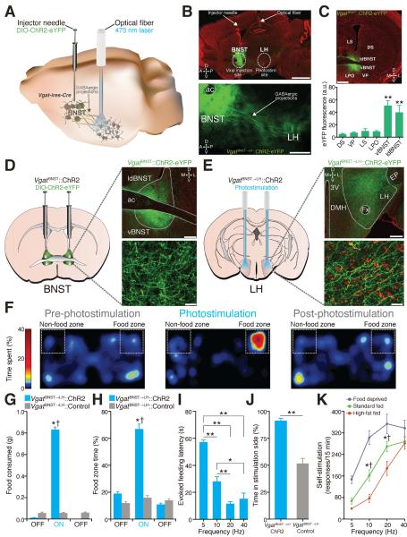
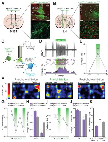
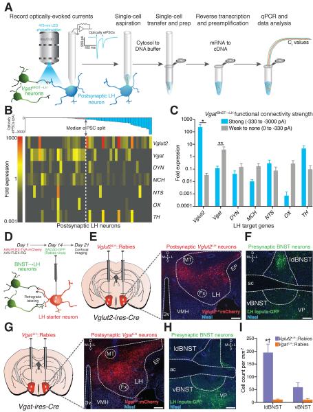
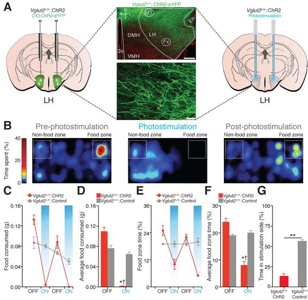
The growing prevalence of overeating disorders is a key contributor to the worldwide obesity epidemic. Dysfunction of particular neural circuits may trigger deviations from adaptive feeding behaviors. The lateral hypothalamus (LH) is a crucial neural substrate for motivated behavior, including feeding, but the precise functional neurocircuitry that controls LH neuronal activity to engage feeding has not been defined. We observed that inhibitory synaptic inputs from the extended amygdala preferentially innervate and suppress the activity of LH glutamatergic neurons to control food intake. These findings help explain how dysregulated activity at a number of unique nodes can result in a cascading failure within a defined brain network to produce maladaptive feeding.


Figures

Fig. 1
Fig. 1. VgatBNST➝LH circuit activation induces feeding in well-fed mice
(A) VgatBNST➝LH circuit targeting. (B) 10x (top) and 20x (bottom) images of the VgatBNST➝LH::ChR2-eYFP circuit (scale bars = 1 mm (top), 500 μm (bottom)). (C) Localized ChR2-eYFP expression in the BNST (top) and quantified eYFP fluorescence intensity (bottom) is significantly greater in the BNST compared to surrounding regions (F5,29 = 11.22, P < 0.001, n = 5 sections from n = 5 mice; ac, anterior commissure; ldBNST, lateral-dorsal BNST; vBNST, ventral BNST; LS, lateral septum; LPO, lateral preoptic area; VP, ventral pallidum; DS, dorsal striatum; scale bar = 200 μm). (D and E) ChR2-eYFP expression in the BNST (D) and axonal projections in the LH (E) in Vgat-ires-Cre mice (LH, lateral hypothalamus; Fx, fornix; EP, entopeduncular nucleus; DMH, dorsomedial hypothalamus; 3V, third ventricle; D, dorsal; V, ventral; L, lateral; M, medial; green = ChR2-eYFP; red = Nissl stain; scale bars = 200 μm (top), 20 μm (bottom)). (F) Spatial location heat maps in 20 min epochs before, during, and after 20-Hz photostimulation-induced feeding. (G and H) Photostimulation of VgatBNST➝LH projections significantly increased grain-based (standard) food intake (F2,24 = 201.6, P < 0.001) (G) and food zone time (F2,24 = 18.61, P < 0.001, n = 5 mice per group) (H). (I) Higher photostimulation frequencies significantly decreased evoked feeding latencies (F3,56 = 48.89, P < 0.001, n = 9 mice). (J) VgatBNST➝LH::ChR2 mice spent significantly more time in the photostimulation-paired side compared to controls (P < 0.001, n = 5 mice per group). (K) Food-deprived VgatBNST➝LH::ChR2 mice nose poked significantly more for 10- and 20-Hz photostimulation when compared to 2 days of standard fed ad libitum and to 2 days of standard fed ad libitum supplemented with 2 hr of high-fat food exposure before the self-stimulation session (F2,204 = 40.87, P < 0.001, n = 9 mice). All values for all figures represent mean ± s.e.m. * P < 0.05, ** P < 0.001 (Student's t-test or ANOVA followed by Bonferroni post-hoc comparisons, where applicable). Dagger symbol denotes significance compared to all manipulations.
Fig. 2
Fig. 2. VgatBNST➝LH circuit inhibition diminishes feeding in food-deprived mice and is aversive
(A and B) eArch3.0-eYFP expression in the BNST (A) and axonal projections in the LH (B) in Vgat-ires-Cre mice (scale bars = 200 μm (top), 20 μm (bottom)). (C) Schematic for anesthetized in vivo extracellular recordings in the LH. (D) Example trace from a single LH unit (top) and its representative peri-event histogram and raster (bottom). (E) The average firing rate of light-responsive LH units significantly increased during the 5-s photoinhibition trials (F2,12 = 19.52, P < 0.001, n = 5 units from n = 3 mice). (F) Spatial location heat maps in 10 min epochs before, during, and after photoinhibition. (G and H) Photoinhibition of VgatBNST➝LH projections significantly decreased standard food intake (F1,44 = 16.30, P < 0.001) and time spent in the food zone (I and J) (F1,44 = 2.43, P = 0.028, n = 6 mice per group). (K) VgatBNST➝LH::eArch3.0 mice spent significantly less time in the photoinhibition-paired side when compared to controls (P = 0.004, n = 6 mice per group).
Fig. 3
Fig. 3. VgatBNST➝LH projections preferentially target LH glutamatergic neurons
(A) Schematic for ChR2-assisted circuit mapping with single-cell gene expression profiling. (B) Color-coded fold expression of all target genes from all recorded LH neurons (Vglut2, vesicular glutamate transporter-2; Vgat, vesicular GABA transporter; DYN, dynorphin; MCH, melanin-concentrating hormone; NTS, neurotensin; OX, orexin/hypocretin; TH, tyrosine hydroxylase). The average fold expression for Vglut2 was significantly higher in postsynaptic LH neurons that display large optically-evoked inhibitory postsynaptic current amplitudes (strongly innervated) compared to weakly innervated LH neurons (U = 169.0, P = 0.016, n = 6 mice, n = 48 cells). (D) Schematic for modified rabies virus tracing. (E and F) Images from a Vglut2-ires-cre mouse showing FLEX-TVA-mCherry expression in LH glutamatergic neurons (E) and appreciable SADΔG-GFP labeling of BNST neurons (F). (G and H) FLEX-TVA-mCherry expression in LH GABAergic neurons (G) and minimal SADΔG-GFP labeling of BNST neurons (H) (green = SADΔG-GFP; red = FLEX-TVA-mCherry; blue = Nissl stain; scale bars = 200 μm). (I) Significantly more BNST neurons innervate LH glutamatergic neurons compared to LH GABAergic neurons (F1,20 = 38.50, P < 0.001, n = 3 mice per group).
Fig. 4
Fig. 4. Photoactivation of Vglut2LH neurons suppresses feeding in food-deprived mice and is aversive
(A) ChR2-eYFP expression in the LH of a Vglut2-ires-Cre mouse (scale bars = 200 μm (top), 20 μm (bottom)). (B) Spatial location heat maps in 10 min epochs before, during, and after 5-Hz photostimulation. (C and D) Photostimulation of Vglut2LH neurons significantly decreased food intake (F1,36 = 13.31, P < 0.001) and food zone time (E and F) (F1,36 = 13.12, P < 0.001, n = 5 mice per group). (G) Vglut2LH::ChR2 mice spent significantly less time in the photostimulation-paired side when compared to controls (P < 0.001, n = 5 mice per group).

Comment in

References

    1. Hoebel BG, Teitelbaum P. Hypothalamic Control of Feeding and Self-Stimulation. Science. 1962;135:375–377. - PubMed
    1. Delgado JR, Anand BK. Increase of food intake induced by electrical stimulation of the lateral hypothalamus. Am. J. Physiol. 1953;172:162–168. - PubMed
    1. Wise RA. Hypothalamic motivational systems: fixed or plastic neural circuits? Science. 1968;162:377–379. - PubMed
    1. Stanley BG, Ha LH, Spears LC, Dee MG., 2nd Lateral hypothalamic injections of glutamate, kainic acid, D,L-alpha-amino-3-hydroxy-5-methyl isoxazole propionic acid or N-methyl-D-aspartic acid rapidly elicit intense transient eating in rats. Brain Res. 1993;613:88–95. - PubMed
    1. Turenius CI, et al. GABA(A) receptors in the lateral hypothalamus as mediators of satiety and body weight regulation. Brain Res. 2009;1262:16–24. - PubMed

Publication types

MeSH terms