. 2013 Sep 20;8(9):e74863.
doi: 10.1371/journal.pone.0074863. eCollection 2013.

Differential host response, rather than early viral replication efficiency, correlates with pathogenicity caused by influenza viruses

Affiliations

Differential host response, rather than early viral replication efficiency, correlates with pathogenicity caused by influenza viruses

Peter S Askovich et al. PLoS One. .

Abstract

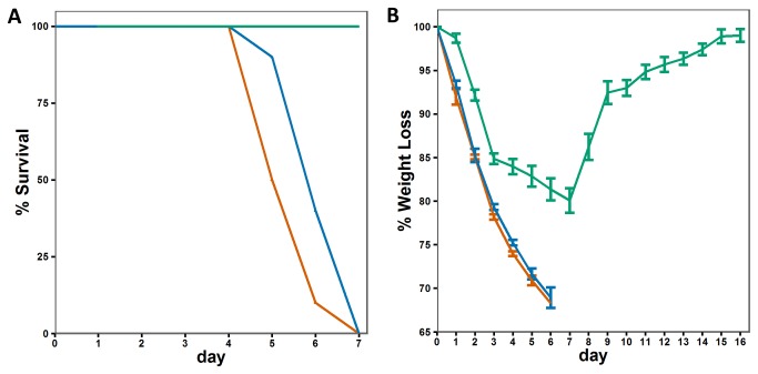
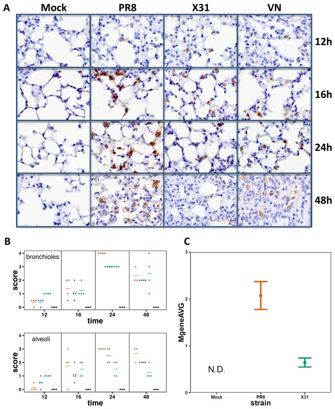
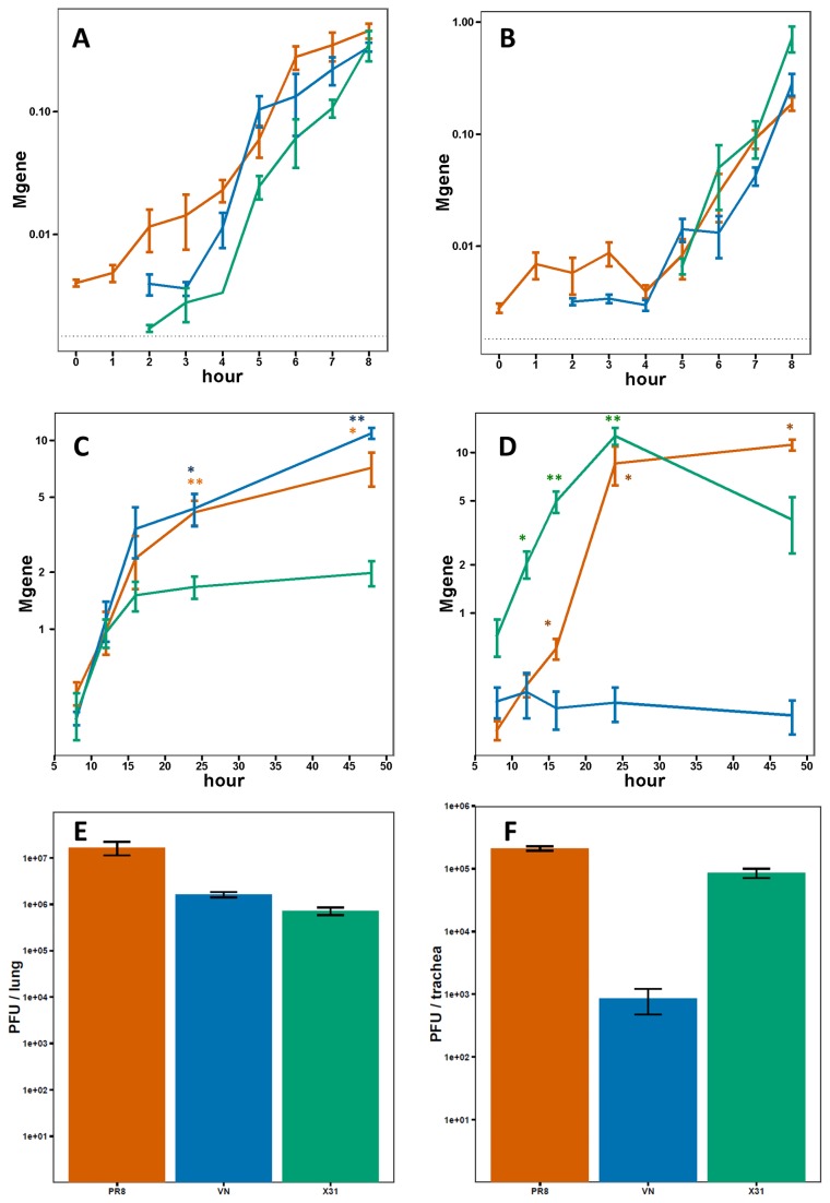
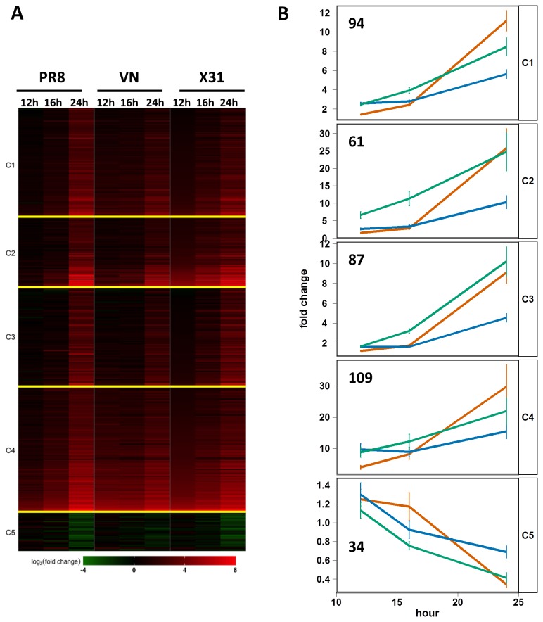
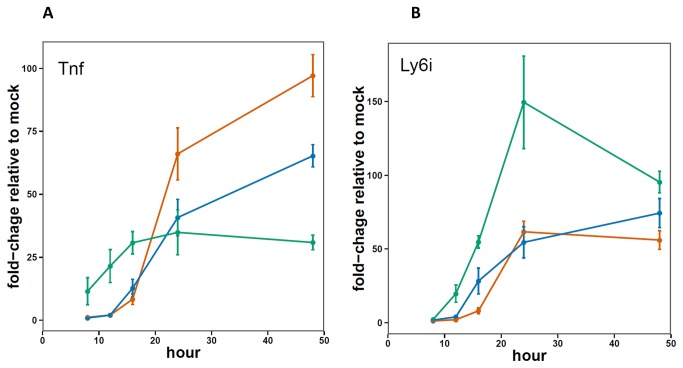
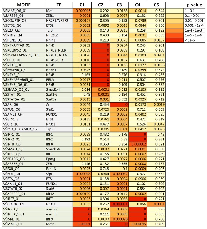
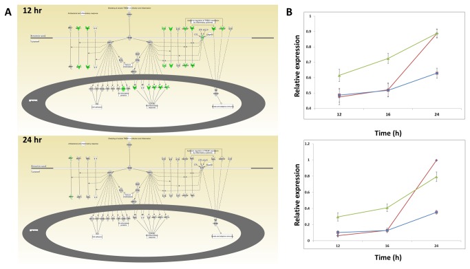
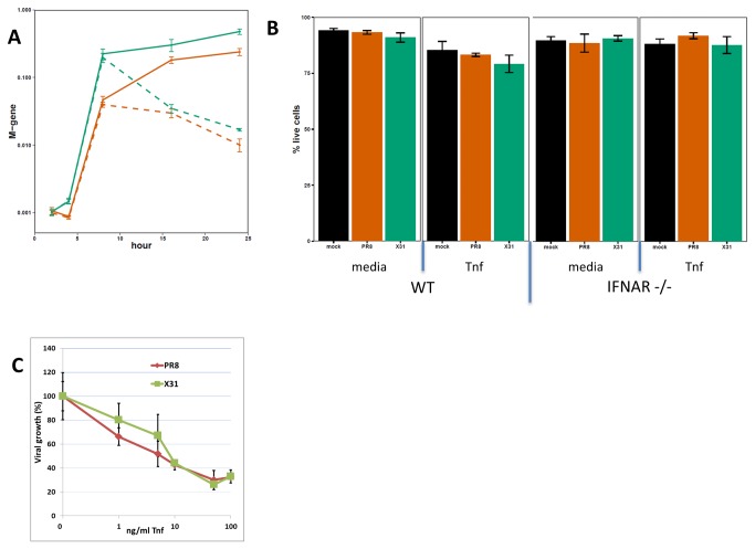
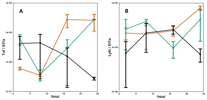
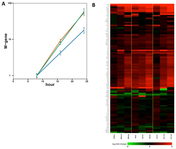
Influenza viruses exhibit large, strain-dependent differences in pathogenicity in mammalian hosts. Although the characteristics of severe disease, including uncontrolled viral replication, infection of the lower airway, and highly inflammatory cytokine responses have been extensively documented, the specific virulence mechanisms that distinguish highly pathogenic strains remain elusive. In this study, we focused on the early events in influenza infection, measuring the growth rate of three strains of varying pathogenicity in the mouse airway epithelium and simultaneously examining the global host transcriptional response over the first 24 hours. Although all strains replicated equally rapidly over the first viral life-cycle, their growth rates in both lung and tracheal tissue strongly diverged at later times, resulting in nearly 10-fold differences in viral load by 24 hours following infection. We identified separate networks of genes in both the lung and tracheal tissues whose rapid up-regulation at early time points by specific strains correlated with a reduced viral replication rate of those strains. The set of early-induced genes in the lung that led to viral growth restriction is enriched for both NF-κB binding site motifs and members of the TREM1 and IL-17 signaling pathways, suggesting that rapid, NF-κB -mediated activation of these pathways may contribute to control of viral replication. Because influenza infection extending into the lung generally results in severe disease, early activation of these pathways may be one factor distinguishing high- and low-pathogenicity strains.


Conflict of interest statement

Competing Interests: The authors have declared that no competing interests exist. Carrie Rosenberger moved to Genentech after completing her part in this project at Seattle BioMed.

Figures

Figure 1
Figure 1. Relative pathogenicity of PR8, X31, and VN.
A) Survival and B) weight-loss for mice infected with 105 PFU (red=PR8, green=X31, blue=VN). Error bars indicate the SEM for 10 mice at each time point.
Figure 2
Figure 2. Replication of influenza virus in mouse lungs.
A) Immunohistochemistry of influenza-infected lungs. Paraffin embedded sections of mouse lungs were stained using an anti-HA antibody. Tissues were collected at the indicated times. B) Histological scoring of the extent and distribution of infection in the lungs. Each dot represents the score for an individual animal; horizontal bars represent the mean score for the group (red=PR8, green=X31, blue=VN). C) Influenza M-gene expression in T1a+ sorted lung cells 18 hours post-infection. T-test p-value for a difference between X31 and PR8 is 0.0018.
Figure 3
Figure 3. Replication of influenza virus in mouse lungs and trachea.
Quantification of viral genomes in the lung and trachea A, B) during the first 8 hours following infection and C,D) from 8 to 48 hours following infection (red=PR8, green=X31, blue=VN). Error bars indicate the SEM for 3-5 mice at each time point. The dotted line indicates the minimum quantifiable level (* p < 0.05, ** p < 0.001 for a non-zero difference in M-gene expression of each strain relative to C). X31 or D) VN). E and F) Viral titer in lungs and trachea of infected mice 24 hours post-infection. Values represent average of 3-5 mice. Error bars show standard error.
Figure 4
Figure 4. Global expression profiling of lung tissue isolated from influenza-infected mice.
A) Heatmap depicting 385 genes whose expression is altered in the lung during the first 24 hours following infection with influenza virus (permutation q-value=<0.001 and > 5-fold difference). B) Mean expression of all genes within each cluster (K means with k=5 for 385 genes) grouped by virus strain (X31 – green; PR8 – red; VN – blue). The number of genes in each cluster is indicated. Error bars depict the SEM for all genes in the cluster at each time point.
Figure 5
Figure 5. Microfluidics qPCR measurements of Tnf and Ly6i expression in the lungs 8-48 hours post infection.
Graphs show average values (red=PR8, green=X31, blue=VN) and error bars depict SEM for 3-5 mice per group.
Figure 6
Figure 6. Transcription factor binding site motifs enriched in at least one expression cluster.
Enriched motifs (p < 10-3) were identified from a Transfac database of 909 unique motifs. Zero (0) indicates motifs enriched at a p-value < 10-6, the limiting resolution of the analysis.
Figure 7
Figure 7. Pathway enrichment analysis.
A) Network diagram of TREM1 signaling canonical pathway from IPA. Colored nodes indicate genes with RNA expression measures above background in murine lungs infected with influenza virus. Green indicates higher expression in X31-infection than PR-infection at the indicated time point (>= 2 Fold). Grey color indicates that the difference in expression is < 2 fold. B) Average expression measure of all genes above background annotated as belonging to the “TREM1 signaling” (top) and “Differential Regulation of Cytokine Production in Macrophages and T Helper Cells by IL-17A and IL-17F” (bottom) pathways. PR8=red, VN=blue, X31=green. Error bars represent the SEM.
Figure 8
Figure 8. PR8 and X31 replication in LET-1 cells.
A) LET-1 cells were infected with either PR8 (red) or X31 (green) and treated with TNF (100 ng/mL) two hours post infection (dashed lines) or buffer only (solid lines). (B) Percent of live LET-1 cells (derived from WT or IFNAR KO mice) at 24h post infection with or without Tnf added to the media (at 2h). (C) Tnf dose response in PR8 and X31 infected LET-1 cells (n=3). Tnf was added 2h post-infection, viral M gene was measured at 24h post-infection. Error bars represent SEM.
Figure 9
Figure 9. Tnf and Ly6i expression in PR8 or X31 infected LET-1 cells.
LET-1 cells were infected with either PR8 (red) or X31 (green) and relative levels of endogenous (A) Tnf and (B) Ly6i transcripts determined from RNA isolated at specified time points. Error bars represent SEM for n=2.
Figure 10
Figure 10. Transcriptional response to influenza infection in mTECs.
A) Expression of the influenza M-gene in mTEC cultures infected with PR8 (red), X31 (green), or VN (blue), at an MOI of 0.01, normalized to expression at 8 hours for each strain. Values show average of 3 replicates and error bars represent the SEM. B) Heatmap depicting expression of 96 genes in mTEC cultures infected with X31, PR8, or VN 8-24h post-infection.

References

    1. WHO (2009) Influenza (Seasonal) Fact Sheet No 211. World Health Organization.
    1. Molinari NA, Ortega-Sanchez IR, Messonnier ML, Thompson WW, Wortley PM et al. (2007) The annual impact of seasonal influenza in the US: measuring disease burden and costs. Vaccine 25: 5086-5096. doi:10.1016/j.vaccine.2007.03.046. PubMed: 17544181. - DOI - PubMed
    1. Meltzer MI, Cox NJ, Fukuda K (1999) The economic impact of pandemic influenza in the United States: priorities for intervention. Emerg Infect Dis 5: 659-671. doi:10.3201/eid0505.990507. PubMed: 10511522. - DOI - PMC - PubMed
    1. Johnson NP, Mueller J (2002) Updating the accounts: global mortality of the 1918-1920 "Spanish" influenza pandemic. Bull Hist Med 76: 105-115. doi:10.1353/bhm.2002.0022. PubMed: 11875246. - DOI - PubMed
    1. Faruqui F, Mukundan D (2010) 2009 pandemic influenza: a review. Curr Opin Pediatr 22: 530-535. doi:10.1097/MOP.0b013e32833bb81a. PubMed: 20601883. - DOI - PubMed

Publication types

MeSH terms