. 2013 Sep 20;8(9):e74992.
doi: 10.1371/journal.pone.0074992. eCollection 2013.

Doublecortin knockout mice show normal hippocampal-dependent memory despite CA3 lamination defects

Affiliations

Doublecortin knockout mice show normal hippocampal-dependent memory despite CA3 lamination defects

Johanne Germain et al. PLoS One. .

Abstract

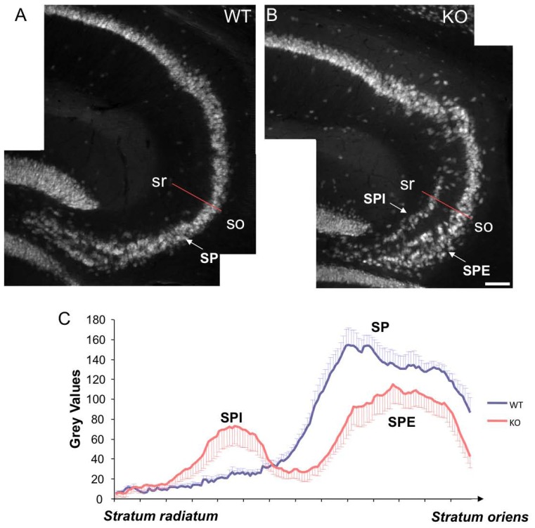
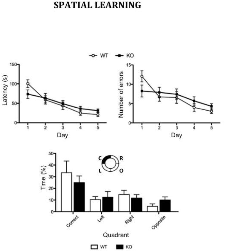
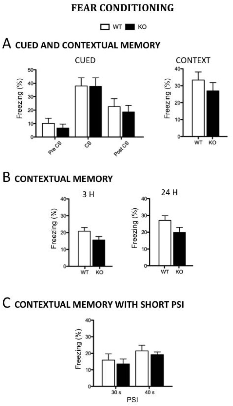
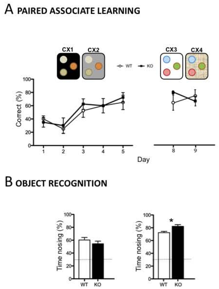
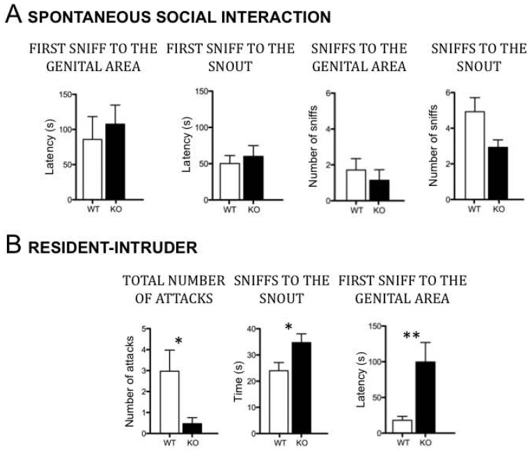
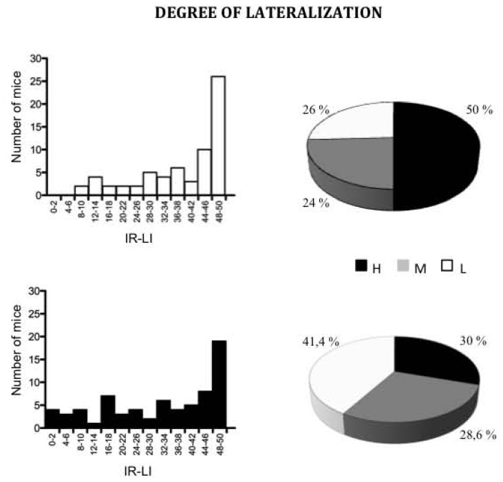
Mutations in the human X-linked doublecortin gene (DCX) cause major neocortical disorganization associated with severe intellectual disability and intractable epilepsy. Although Dcx knockout (KO) mice exhibit normal isocortical development and architecture, they show lamination defects of the hippocampal pyramidal cell layer largely restricted to the CA3 region. Dcx-KO mice also exhibit interneuron abnormalities. As well as the interest of testing their general neurocognitive profile, Dcx-KO mice also provide a relatively unique model to assess the effects of a disorganized CA3 region on learning and memory. Based on its prominent anatomical and physiological features, the CA3 region is believed to contribute to rapid encoding of novel information, formation and storage of arbitrary associations, novelty detection, and short-term memory. We report here that Dcx-KO adult males exhibit remarkably preserved hippocampal- and CA3-dependant cognitive processes using a large battery of classical hippocampus related tests such as the Barnes maze, contextual fear conditioning, paired associate learning and object recognition. In addition, we show that hippocampal adult neurogenesis, in terms of proliferation, survival and differentiation of granule cells, is also remarkably preserved in Dcx-KO mice. In contrast, following social deprivation, Dcx-KO mice exhibit impaired social interaction and reduced aggressive behaviors. In addition, Dcx-KO mice show reduced behavioral lateralization. The Dcx-KO model thus reinforces the association of neuropsychiatric behavioral impairments with mouse models of intellectual disability.


Conflict of interest statement

Competing Interests: The authors have declared that no competing interests exist.

Figures

Figure 1
Figure 1. Abnormal hippocampal lamination in the Dcx-KO mice.
(A–B) NeuN labeled sagittal sections of adult hippocampus from a control (A) and a Dcx-KO (B) mouse showing a largely normal CA1 region but a disorganized CA3 region. This disorganization is characterized by two distinct pyramidal cell layers (termed stratum pyramidale internal, SPI, and stratum pyramidale external, SPE). (C) Histogram showing the distribution of pixel intensities along the red lines as indicated in A and B, across the CA3 region and extending from the stratum radiatum (sr) towards the stratum oriens (so) from both control (blue) and KO (red) mice. The distribution of these grey values clearly shows a double peak in the KOs corresponding to SPI and SPE respectively, in comparison to a single peak (corresponding to the stratum pyramidal, SP) in the WTs. Scale bar 60 µm.
Figure 2
Figure 2. Normal spatial learning and memory in the Dcx-KO mice.
During acquisition trials in the Barnes maze, performances of Dcx-KO (n = 7) and WT (n = 8) male mice are expressed as latency (s) to escape the platform and number of errors. During the probe test, spatial strategy is expressed as percentage of time spent at the periphery of the maze, in the target, right, left, and opposite quadrants, as indicated in the schema. Values represent means +/− SEM.
Figure 3
Figure 3. Normal fear conditioning in the Dcx-KO mice. A/Cued and contextual memory;
percentage of time spent freezing to tone (left) and to context (right) 24h after training in two context-tone-shock pairing trials (n = 8–9 mice per group); B/Contextual memory ; percentage of time spent freezing to context 3 h (left) (n = 13 mice per group) and 24 h (right) after training in one context/shock pairing trial (n = 9–16 mice per group); C/Contextual memory ; percentage of time spent freezing following a short PSI (30 s: n = 6–8 mice per group; 40 s: n = 19–21 mice per group). Values represent means +/− SEM.
Figure 4
Figure 4. Normal contingent learning and novelty detection in the Dcx-KO mice. A/Paired-associate learning;
performances of Dcx-KO and WT (n = 4 mice per genotype) male mice trained for five days in the paired-associate learning between two contexts (CX1 and CX2) and three olfactory cues, one baited (+) and two unbaited (−), followed by two days using novel contexts and novel odors. B/Novelty detection; Performances of Dcx-KO male mice expressed as the percentage of time spent exploring the displaced object (left) (n = 15 mice per genotype) or the novel object (right) (n = 10 mice per genotype) three minutes after the last habituation trials. The horizontal line indicates the by-chance exploration time for each of the three objects (33%). Values represent means +/− SEM.
Figure 5
Figure 5. Abnormal social interaction and reduced aggressive behavior in the Dcx-KO mice. A/Spontaneous social interaction
; Performances of Dcx-KO male mice (n = 7 mice per genotype) expressed as the latency of the first sniff to the genital area or snout, and the total amount of time spent sniffing the genital area or the snout, measured over a 5-min period. B/Resident-Intruder ; Performances of Dcx-KO (n = 17) and WT (n = 16) male mice in the resident-intruder test expressed the number of attacks, the total amount of time spent sniffing the snout, and the latency of the first anogenital sniff, and, measured over a 10-min period. Values represent means +/− SEM. *p<0.05, **p<0.01.
Figure 6
Figure 6. Reduced behavioral lateralization in the Dcx-KO mice.
Degree of lateralization in Dcx-KO (n = 70) and WT (n = 66) male mice. A/Distribution for the |R–L| variable that takes even values from 0 to 50; B/Distributions for the three classes of highly (H) lateralized mice (|R–L| ≥46), ambidextrous (L) mice (|R–L| ≤30), and intermediate (M) mice scoring between 32–44.

Similar articles

Cited by

References

    1. Kappeler C, Dhenain M, Phan Dinh Tuy F, Saillour Y, Marty S, et al. (2007) Magnetic resonance imaging and histological studies of corpus callosal and hippocampal abnormalities linked to doublecortin deficiency. J Comp Neurol 500: 239–254. - PubMed
    1. Gleeson JG, Allen KM, Fox JW, Lamperti ED, Berkovic S, et al. (1998) Doublecortin, a brain-specific gene mutated in human X-linked lissencephaly and double cortex syndrome, encodes a putative signaling protein. Cell 92: 63–72. - PubMed
    1. Hong SE, Shugart YY, Huang DT, Shahwan SA, Grant PE, et al. (2000) Autosomal recessive lissencephaly with cerebellar hypoplasia is associated with human RELN mutations. Nat Genet 26: 93–96. - PubMed
    1. Keays DA, Tian G, Poirier K, Huang GJ, Siebold C, et al. (2007) Mutations in alpha-tubulin cause abnormal neuronal migration in mice and lissencephaly in humans. Cell 128: 45–57. - PMC - PubMed
    1. Reiner O, Carrozzo R, Shen Y, Wehnert M, Faustinella F, et al. (1993) Isolation of a Miller-Dieker lissencephaly gene containing G protein beta-subunit-like repeats. Nature 364: 717–721. - PubMed

Publication types