. 2013 Sep 20;8(9):e75002.
doi: 10.1371/journal.pone.0075002. eCollection 2013.

Orthopedia transcription factor otpa and otpb paralogous genes function during dopaminergic and neuroendocrine cell specification in larval zebrafish

Affiliations

Orthopedia transcription factor otpa and otpb paralogous genes function during dopaminergic and neuroendocrine cell specification in larval zebrafish

António M Fernandes et al. PLoS One. .

Abstract

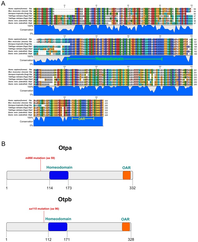
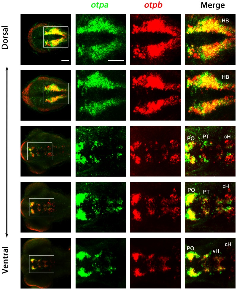
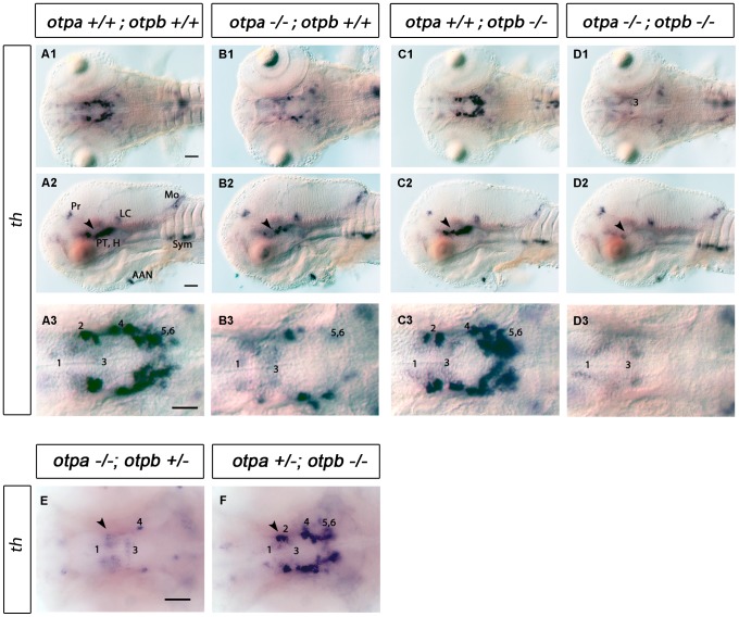
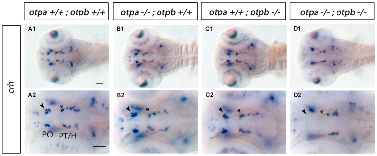
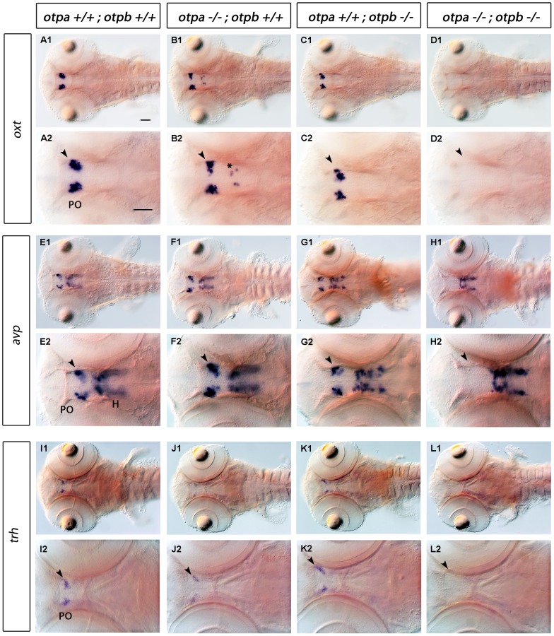
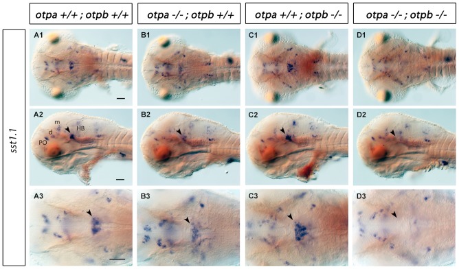
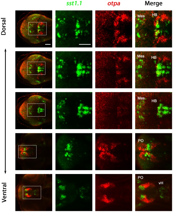
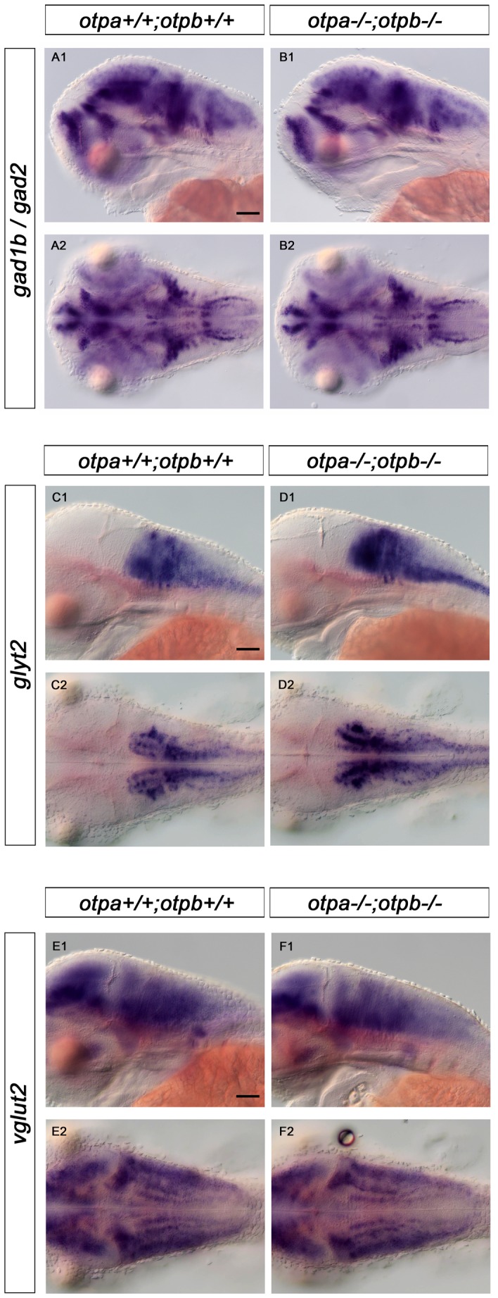
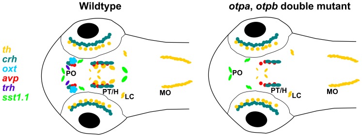
The homeodomain transcription factor Orthopedia (Otp) is an important regulator for specification of defined subsets of neuroendocrine cells and dopaminergic neurons in vertebrates. In zebrafish, two paralogous otp genes, otpa and otpb, are present in the genome. Neither complete loss of Otp activity nor differential contributions of Otpa and Otpb to specification of defined neuronal populations have been analyzed in detail. We characterized zebrafish embryos and early larvae mutant for null alleles of otpa, otpb, or both genes to determine their individual contributions to the specification of th expressing dopaminergic neuronal populations as well as of crh, oxt, avp, trh or sst1.1 expressing neuroendocrine cells. otpa mutant larvae show an almost complete reduction of ventral diencephalic dopaminergic neurons, as reported previously. A small reduction in the number of trh cells in the preoptic region is detectable in otpa mutants, but no significant loss of crh, oxt and avp preoptic neuroendocrine cells. otpb single mutant larvae do not display a reduction in dopaminergic neurons or neuroendocrine cells in the otp expressing regions. In contrast, in otpa and otpb double mutant larvae specific groups of dopaminergic neurons as well as of crh, oxt, avp, trh and sst1.1-expressing neuroendocrine cells are completely lost. These observations suggest that the requirement for otpa and otpb function during development of the larval diencephalon is partially redundant. During evolutionary diversification of the paralogous otp genes, otpa maintained the prominent role in ventral diencephalic dopaminergic and neuroendocrine cell specification and is capable of partially compensating otpb loss of function. In addition, we identified a role of Otp in the development of a domain of somatostatin1-expressing cells in the rostral hindbrain, a region with strong otp expression but so far uncharacterized Otp function. Otp may thus be crucial for defined neuronal cell types also in the hindbrain.


Conflict of interest statement

Competing Interests: The authors have declared that no competing interests exist.

Figures

Figure 1
Figure 1. Otpbsa0115 mutants lack the highly conserved Homeodomain.
(A) The Otp protein and specifically the homeodomain sequence (green label) are highly conserved in vertebrates. The conserved OAR domain present in all Otp proteins in vertebrates is also depicted (yellow label). (B) Schematic representation showing both Otpa and Otpb protein structure and position of stop codons caused by mutations. In the otpam866 allele the mutation results in a frameshift and a premature stop codon after additional 59 amino acids. In the otpbsa0115 allele a base-pair deletion results in a premature stop codon 96 amino acids downstream of the start codon. Both mutations generate smaller proteins which completely lack the highly conserved homeodomain (in blue). The conserved OAR (otp, aristaless, rax), potential interaction domain present in several paired-like homeodomain proteins and all Otp proteins in vertebrates is also depicted (orange).
Figure 2
Figure 2. Expression of otpa and otpb in wildtype larvae.
Expression of otpa and otpb were detected by double fluorescent whole mount in situ hybridization of wildtype larvae fixed at 3 dpf. From the whole confocal image stack, sub-stacks ranging from dorsal hindbrain image planes to ventral forebrain planes were used to generate a series of dorso-ventral Z-projections. The data reveal that otpa and otpb have overlapping expression but also non-overlapping domains. Dorsal view, anterior at left. Abbreviations: cH, caudal hypothalamus; HB, hindbrain; PO, preoptic region; PT, posterior tuberculum; vH, ventral hypothalamus. Scale bar is 50 µm.
Figure 3
Figure 3. Analysis of DA neurons by expression of th in otpa and otpb single and double mutant larvae.
(A–D) Whole-mount in situ hybridization of 3 dpf larvae reveals reduction of th expression in the posterior tuberculum of otpa and total loss of the expression in otpa;otpb double mutants (arrowhead). Other th expressing domains are not affected. (A1–D1, A3–D3) Dorsal views, anterior at left; (A2–D2) lateral views, dorsal up. Scale bar is 50 µm. (E,F) Whole-mount in situ hybridization of 3 dpf larvae reveals reduction of th expression in the posterior tuberculum of otpa mutant, otpb heterozygous larvae (E) (arrowhead). No clear reduction is detected in the posterior tuberculum of otpb mutant, otpa heterozygous larvae (F) (arrowhead). Dorsal view, anterior at left. Scale bar is 50 µm. Abbreviations: AAC, arch associated cluster; DC, diencephalic cluster; H, hypothalamus; LC, locus coeruleus; MO, medulla oblongata; Pr, pretectum; PT, posterior tuberculum. Numbers indicate dopaminergic neurons in the ventral thalamic cluster (1) and posterior tuberculum/hypothalamus (2–6) according to . Scale bar is 50 µm.
Figure 4
Figure 4. Expression of crh in otpa and otpb single and double mutant larvae.
Whole-mount in situ hybridization of 3 dpf larvae reveals loss of crh expression in the preoptic region and posterior tuberculum of otpa;otpb double mutant larvae (arrowhead and asterisk, respectively). Dorsal view, anterior at left. Scale bar is 50 µm. Abbreviations: H, hypothalamus; PO, preoptic region; PT, posterior tuberculum.
Figure 5
Figure 5. Expression of oxt, avp and trh in otpa and otpb single and double mutant larvae.
Whole-mount in situ hybridization reveals loss of oxt, avp and trh expression in the preoptic region (arrowhead) of otpa;otpb double mutant larvae at 3 dpf. We detected oxt-expressing cells at ectopic locations within the diencephalon in otpa mutants (B2, asterisk). A reduction of trh-expressing cells in the preoptic region in otpa single mutants is detectable, (J2, arrowhead). Dorsal view, anterior at left. Scale bar is 50 µm. Abbreviations: H, hypothalamus; PO, preoptic region.
Figure 6
Figure 6. Expression of sst1.1 in otpa and otpb single and double mutant larvae.
Whole-mount in situ hybridization reveals reduction of sst1.1 expression (arrowhead) in the rostral hindbrain of otpa mutants and total loss of the expression in otpa;otpb double mutant larvae at 3 dpf. (A1–D1, A3–D3) Dorsal view, anterior at left; (A2–D2) lateral view, dorsal up. Scale bar is 50 µm. Abbreviations: d, diencephalon; HB, hindbrain; m, mesencephalon; PO, preoptic region.
Figure 7
Figure 7. Analysis of coexpression of otpa and sst1.1 in wildtype larvae.
Expression of otpa and sst1.1 were detected by double fluorescent whole mount in situ hybridization of wildtype larvae fixed at 3 dpf. From the whole confocal image stack, sub-stacks ranging from dorsal hindbrain image planes to ventral forebrain planes were used to generate a series of dorso-ventral Z-projections. The data reveal that sst1.1 and otpa expression domains overlap in the rostral hindbrain in wildtype larvae at 3 dpf, and some cells appear to coexpress both genes. Dorsal view, anterior at left. Abbreviations: cH, caudal hypothalamus; HB, hindbrain; Mes, mesencephalon; PO, preoptic region; vH, ventral hypothalamus. Scale bar is 50 µm.
Figure 8
Figure 8. Expression of otpa in relation to gabaergic, glutamatergic and glycinergic markers in the hindbrain of wildtype larvae.
Potential coexpression of otpa with gabaergic (gad2, A), glutamatergic (vglut2, B) and glycinergic (glyt2, C) markers was analyzed by double whole mount FISH at 3 dpf. A1, B1, and C1 are single plane dorsal views of the hindbrain, anterior is to the left. A2, B2, and C2 are cross-sections at the level of the hindbrain indicated by the white line in A1, B1, and C1, respectively. The orthogonal view cross sections were obtained from dorsal confocal stacks using the TransformJ Turn plugin of the ImageJ software. Scale bar is 50 µm.
Figure 9
Figure 9. Expression of gad1b/2, glyt2 and vglut2 in wildtype and in otp;otpb double mutant larvae.
Whole-mount in situ hybridization reveals no changes in expression of gabaergic (gad1b/gad2), glycinergic (glyt2)and glutamatergic (vglut2) in the hindbrain of otpa;otpb double mutant larvae at 3 dpf. (A1, B1, C1, D1, E1, F1) lateral view, dorsal up; (A2, B2, C2, D2, E2,F2) dorsal view, anterior at left. Scale bar is 50 µm.
Figure 10
Figure 10. Schematic representation of neuroendocrine and dopaminergic cell groups affected in otpa/otpb double mutant larval zebrafish at 3 dpf.
Schematic diagram showing the expression of the th, sst1.1, trh, oxt, avp, and crh neuronal groups analyzed in this study at 3 dpf in zebrafish larvae. For simplicity, the schematic representation does not include the entire expression patterns of all these 7 genes, but focuses on anatomical regions with Otp expression. The comparison of wildtype and otpa;otpb double mutant larvae reveals groups dependent on Otp activity. H, hypothalamus; LC, locus coeruleus; MO, medulla oblongata; PO, preoptic region; PT, posterior tuberculum.

References

    1. Simeone A, D’Apice MR, Nigro V, Casanova J, Graziani F, et al. (1994) Orthopedia, a novel homeobox-containing gene expressed in the developing CNS of both mouse and Drosophila. Neuron 13: 83–101. - PubMed
    1. Acampora D, Postiglione MP, Avantaggiato V, Di BM, Vaccarino FM, et al. (1999) Progressive impairment of developing neuroendocrine cell lineages in the hypothalamus of mice lacking the Orthopedia gene. Genes Dev 13: 2787–2800. - PMC - PubMed
    1. Wang W, Lufkin T (2000) The murine Otp homeobox gene plays an essential role in the specification of neuronal cell lineages in the developing hypothalamus. Dev Biol 227: 432–449. - PubMed
    1. Bardet SM, Martinez-de-la-Torre M, Northcutt RG, Rubenstein JL, Puelles L (2008) Conserved pattern of OTP-positive cells in the paraventricular nucleus and other hypothalamic sites of tetrapods. Brain Res Bull 75: 231–235. - PubMed
    1. Moret F, Christiaen L, Deyts C, Blin M, Vernier P, et al. (2005) Regulatory gene expressions in the ascidian ventral sensory vesicle: evolutionary relationships with the vertebrate hypothalamus. Dev Biol 277: 567–579. - PubMed

Publication types

MeSH terms

LinkOut - more resources