Comparative Study
. 2013 Sep 20;8(9):e75836.
doi: 10.1371/journal.pone.0075836. eCollection 2013.

Matrix metalloproteinase-10/TIMP-2 structure and analyses define conserved core interactions and diverse exosite interactions in MMP/TIMP complexes

Affiliations
Comparative Study

Matrix metalloproteinase-10/TIMP-2 structure and analyses define conserved core interactions and diverse exosite interactions in MMP/TIMP complexes

Jyotica Batra et al. PLoS One. .

Abstract

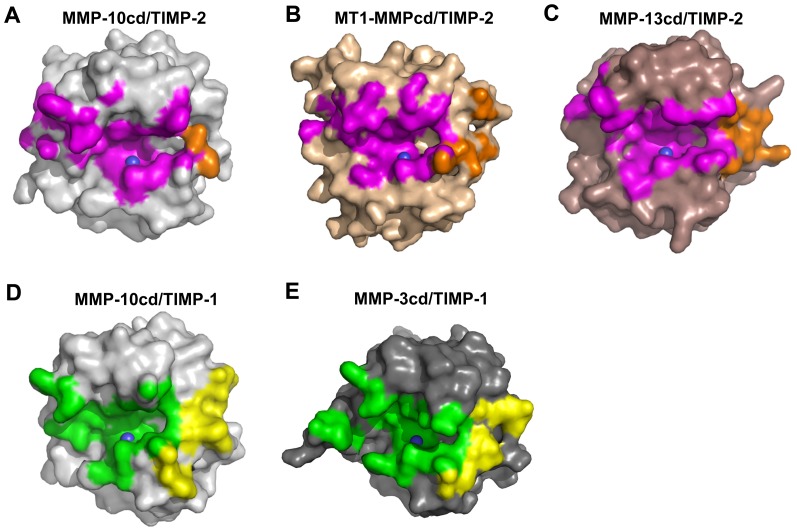
Matrix metalloproteinases (MMPs) play central roles in vertebrate tissue development, remodeling, and repair. The endogenous tissue inhibitors of metalloproteinases (TIMPs) regulate proteolytic activity by binding tightly to the MMP active site. While each of the four TIMPs can inhibit most MMPs, binding data reveal tremendous heterogeneity in affinities of different TIMP/MMP pairs, and the structural features that differentiate stronger from weaker complexes are poorly understood. Here we report the crystal structure of the comparatively weakly bound human MMP-10/TIMP-2 complex at 2.1 Å resolution. Comparison with previously reported structures of MMP-3/TIMP-1, MT1-MMP/TIMP-2, MMP-13/TIMP-2, and MMP-10/TIMP-1 complexes offers insights into the structural basis of binding selectivity. Our analyses identify a group of highly conserved contacts at the heart of MMP/TIMP complexes that define the conserved mechanism of inhibition, as well as a second category of diverse adventitious contacts at the periphery of the interfaces. The AB loop of the TIMP N-terminal domain and the contact loops of the TIMP C-terminal domain form highly variable peripheral contacts that can be considered as separate exosite interactions. In some complexes these exosite contacts are extensive, while in other complexes the AB loop or C-terminal domain contacts are greatly reduced and appear to contribute little to complex stability. Our data suggest that exosite interactions can enhance MMP/TIMP binding, although in the relatively weakly bound MMP-10/TIMP-2 complex they are not well optimized to do so. Formation of highly variable exosite interactions may provide a general mechanism by which TIMPs are fine-tuned for distinct regulatory roles in biology.


Conflict of interest statement

Competing Interests: The authors have declared that no competing interests exist.

Figures

Figure 1
Figure 1. Crystal structure of MMP-10cd/TIMP-2 complex.
(A) Structural overview of the complex shows the MMP-10cd in blue, zinc ions in yellow, calcium ions in orange, disulfide bridges in gold, and TIMP-2 in raspberry. The N-terminal domain of TIMP-2 is positioned to the left, comprising β-strands A–F, helices 1–3 and intervening loops, and the C-terminal domain is positioned on the right, comprising β-strands G–J, helices 4a and 4b, and connecting loops. Numbering shown is consistent with early descriptions of TIMP-1 and TIMP-2 structures from the Bode group , ; helix 4a within the multiple turn loop was originally not numbered. (B) A closer view of the cartoon structure in the vicinity of the active site shows the TIMP-2 N-terminal residue Cys-1 (red arrow) coordinated to the catalytic zinc ion directly behind. (C) The 2Fo-Fc electron density map contoured at 2.0σ around the MMP-10cd active site shows the catalytic zinc coordinated by side chains of His-217, Glu-218, His-221, and His-227. The bound molecule of TIMP-2 also coordinates the catalytic zinc via the terminal amine and carbonyl oxygen of Cys-1. Numbers shown in blue correspond to MMP-10 residues and numbers in red to TIMP-2 residues.
Figure 2
Figure 2. Structural adaptations induced by MMP-10cd/TIMP-2 association.
(A) Structural differences in TIMP-2 bound to the MMP-10cd relative to the crystal structure of unbound TIMP-2 (PDB ID: 1BR9 , shown in wheat) are confined to several surface loops, most notably the AB, C-connector-D, GH, and IJ loops, highlighted by red boxes. (B) Structural alterations in MMP-10cd relative to the crystal structure of MMP-10cd bound to small molecule inhibitor NNGH (PDB ID: 1Q3A , shown in white) are limited to the specificity loop, highlighted by the red box. Superpositions are based on the Cα atoms of all corresponding residues.
Figure 3
Figure 3. Contacts at the MMP-10cd/TIMP-2 interface.
MMP-10cd is rendered as a cartoon covered by semitransparent surface (slate) in the standard frontal orientation, with horizontally aligned TIMP-2 segments in stick representation (salmon). (A) Overview shows contacts of the TIMP-2 C-connector and N-terminal segment with MMP-10 substrate binding cleft (center), TIMP-2 AB loop contacts with MMP-10 S-loop and βIV-βV loop (upper left), and TIMP-2 C-terminal domain contacts with MMP-10 specificity and βV-hB loops (lower right). (B) Closer view of MMP-10 substrate binding cleft shows TIMP-2 C-connector residues occupying nonprimed subsites to the left of the catalytic zinc, while TIMP-2 N-terminal residues occupy primed subsites to the right of the zinc. (C) Closer view of the AB loop interactions reveals two interfacial H-bonds (dotted yellow lines highlighted by yellow arrows), and burial of the Ile-40 side chain in a hydrophobic pocket formed by MMP-10 residues Phe-170, Tyr-171, and Leu190. (D) GH loop residues 132–135 and multiple turn loop residues 151–157 on the C-terminal domain of TIMP-2 form minimal interactions with the MMP-10cd, including ring-stacking and cation-π interactions with MMP-10 specificity loop residues Phe-242 and Tyr-239.
Figure 4
Figure 4. Comparison of MMP-10cd/TIMP-1 and MMP-10cd/TIMP-2 complexes.
MMP-10cd/TIMP-2 molecules are shown in blue and raspberry, respectively, with MMP-10cd/TIMP-1 complex (PDB ID: 3V96) shown in white; complexes are superposed based on Cα atoms of all MMP-10cd residues. (A) The long AB loop of TIMP-2 forms a much more extensive contact area with the MMP-10cd than is seen with TIMP-1, while the C-terminal loops of TIMP-2 form fewer contacts than in the complex with TIMP-1. (B) TIMP-2 is rotated by ∼21° around an axis centered on the catalytic zinc when compared with TIMP-1.
Figure 5
Figure 5. MMP/TIMP complexes feature conserved core interactions but highly diverse peripheral interactions.
MMP-10cd/TIMP-2 (indigo/raspberry) is superposed with four different MMPcd/TIMP structures based on Cα atoms of all corresponding MMP residues: MMP-10cd/TIMP-1 (slate/chartreuse; PDB ID: 3V96) , MMP-3cd/TIMP-1 (purple/yellow; PDB ID: 1UEA) , MT1-MMPcd/TIMP-2 (cyan/brown; PDB ID: 1BQQ) , and MMP-13cd/TIMP-2 (forest/orange; PDB ID: 2E2D) . (A) Positioning of peripheral TIMP loops including the AB and GH loops relative to the MMP show wide variability. (B) In the MMP active site, backbone positioning of TIMP residues 1–4 and the C-connector loop are nearly identical.
Figure 6
Figure 6. Diversity of MMP contact surfaces interacting with TIMP N- and C-terminal domains.
MMPs are shown in the standard frontal orientation, with catalytic zinc shown as a blue sphere. (A) MMP-10cd/TIMP-2: MMP-10cd surface is shown in grey with footprint of the surface buried by TIMP-2 N-terminal domain in magenta and C-terminal domain in orange. (B) MT1-MMPcd/TIMP-2 (PDB ID: 1BQQ) : MT1-MMPcd surface is shown in wheat with footprint of the surface buried by TIMP-2 N-terminal domain in magenta and C-terminal domain in orange. (C) MMP-13cd/TIMP-2 (PDB ID: 2E2D) : MMP-13cd surface is shown in brown with footprint of the surface buried by TIMP-2 N-terminal domain in magenta and C-terminal domain in orange. (D) MMP-10cd/TIMP-1 (PDB ID: 3V96) : MMP-10cd surface is shown in grey with footprint of the surface buried by TIMP-1 N-terminal domain in green and C-terminal domain in yellow. (E) MMP-3cd/TIMP-1 (PDB ID: 1UEA) : MMP-3cd surface is shown in dark grey with footprint of the surface buried by TIMP-1 N-terminal domain in green and C-terminal domain in yellow. The MMP contact surfaces shown are for atoms within 4.5 Å of the TIMP; figures were generated using PYMOL.

References

    1. Stamenkovic I (2003) Extracellular matrix remodelling: the role of matrix metalloproteinases. J Pathol 200: 448–464. - PubMed
    1. Parks WC, Wilson CL, Lopez-Boado YS (2004) Matrix metalloproteinases as modulators of inflammation and innate immunity. Nat Rev Immunol 4: 617–629. - PubMed
    1. Van Wart HE, Birkedal-Hansen H (1990) The cysteine switch: a principle of regulation of metalloproteinase activity with potential applicability to the entire matrix metalloproteinase gene family. Proc Natl Acad Sci U S A 87: 5578–5582. - PMC - PubMed
    1. Rosenblum G, Van den Steen PE, Cohen SR, Grossmann JG, Frenkel J, et al. (2007) Insights into the structure and domain flexibility of full-length pro-matrix metalloproteinase-9/gelatinase B. Structure. 15: 1227–1236. - PubMed
    1. Brew K, Nagase H (2010) The tissue inhibitors of metalloproteinases (TIMPs): an ancient family with structural and functional diversity. Biochim Biophys Acta 1803: 55–71. - PMC - PubMed

Publication types

MeSH terms

Substances