. 2013 Jun 25:2:862-72.
doi: 10.1016/j.nicl.2013.06.011. eCollection 2013.

Impaired and facilitated functional networks in temporal lobe epilepsy

Affiliations

Impaired and facilitated functional networks in temporal lobe epilepsy

Luigi Maccotta et al. Neuroimage Clin. .

Abstract

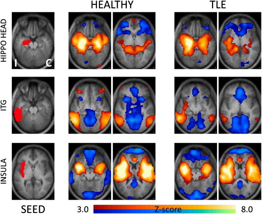
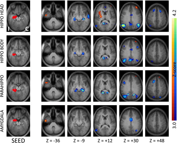
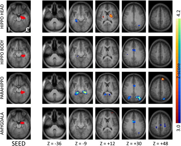
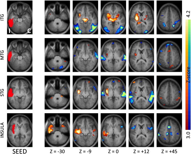
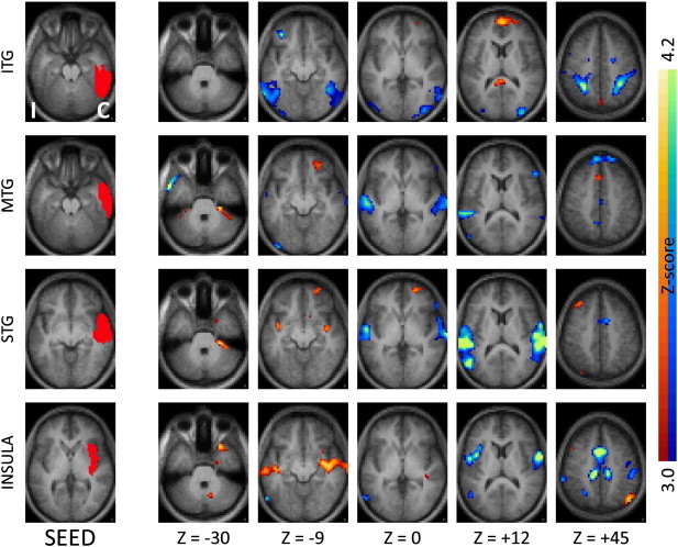
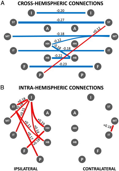
How epilepsy affects brain functional networks remains poorly understood. Here we investigated resting state functional connectivity of the temporal region in temporal lobe epilepsy. Thirty-two patients with unilateral temporal lobe epilepsy underwent resting state blood-oxygenation level dependent functional magnetic resonance imaging. We defined regions of interest a priori focusing on structures involved, either structurally or metabolically, in temporal lobe epilepsy. These structures were identified in each patient based on their individual anatomy. Our principal findings are decreased local and inter-hemispheric functional connectivity and increased intra-hemispheric functional connectivity ipsilateral to the seizure focus compared to normal controls. Specifically, several regions in the affected temporal lobe showed increased functional coupling with the ipsilateral insula and immediately neighboring subcortical regions. Additionally there was significantly decreased functional connectivity between regions in the affected temporal lobe and their contralateral homologous counterparts. Intriguingly, decreased local and inter-hemispheric connectivity was not limited or even maximal for the hippocampus or medial temporal region, which is the typical seizure onset region. Rather it also involved several regions in temporal neo-cortex, while also retaining specificity, with neighboring regions such as the amygdala remaining unaffected. These findings support a view of temporal lobe epilepsy as a disease of a complex functional network, with alterations that extend well beyond the seizure onset area, and the specificity of the observed connectivity changes suggests the possibility of a functional imaging biomarker for temporal lobe epilepsy.

Keywords: Epilepsy; Functional connectivity; Hippocampus; Insula; Temporal lobe; fMRI.


Figures

Supplementary Fig. 1.
Seed-based correlation maps showing the group main effect for healthy controls and TLE patients. Representative seeds in the ipsilateral medial temporal (hippocampal head), lateral temporal (ITG) and insular regions are shown in the left column. The remaining columns show the Z-score maps at selected axial locations. As in prior figures, the hemisphere ipsilateral to the seizure focus is mapped on the left (I) while the contralateral hemisphere is mapped on the right (C). Illustrated clusters are significant (p < .01) after correction for multiple comparisons. Blue–green colors indicate negative correlations, red–yellow indicate positive correlations.
Fig. 1
Fig. 1
Seed-based correlation maps comparing TLE patients to controls for seeds in ipsilateral medial temporal regions. The left column shows the seed, while the remaining columns show the Z-score maps at selected axial slices. Slice labels are in Talairach coordinates (Talairach and Tournoux, 1988). The hemisphere ipsilateral to the seizure focus is mapped on the left (“I”) while the contralateral hemisphere is mapped on the right (“C”). Illustrated clusters are significant (p < .01) after correction for multiple comparisons. Blue–green colors indicate decreased correlations in patients, red–yellow indicate increased correlations. Ipsilateral hippocampal head, body and parahippocampus but not amygdala show significant functional decoupling with their contralateral homologues in TLE patients. The ipsilateral hippocampal head also shows increased functional coupling with the ipsilateral insula.
Fig. 2
Fig. 2
Seed-based correlation maps as in Fig. 1 comparing TLE patients to controls for seeds in contralateral medial temporal regions. Note the decreased functional connectivity with the ipsilateral medial temporal regions, mirroring the effects seen with the ipsilateral seeds.
Fig. 3
Fig. 3
Seed-based correlation maps as in Fig. 1 for seeds in ipsilateral neocortical temporal regions and insula. Note the significantly reduced inter-hemispheric correlations, and significantly increased coupling with the ipsilateral insula and immediately neighboring subcortical regions. ITG — inferior temporal gyrus, MTG — middle temporal gyrus, STG — superior temporal gyrus.
Fig. 4
Fig. 4
Seed-based correlation maps as in Fig. 1 comparing TLE patients to controls for seeds in the contralateral neocortical temporal regions and insula. Note the decreased functional connections with the ipsilateral lateral temporal regions, mirroring the effects seen with the ipsilateral seeds.
Fig. 5
Fig. 5
Network map of correlation differences between TLE patients and controls — A: cross-hemispheric connections; B: intra-hemispheric connections. Blue lines represent significantly decreased correlations in patients compared to controls; red lines represent significantly increased correlations in patients compared to controls (p < .01). Strength of the difference in coupling between patients and controls is indicated numerically next to each connecting line as difference between correlation strengths as well as by the relative line thickness. Note significant functional decoupling across hemispheres that affects both medial and lateral/neocortical temporal regions. Note increased correlations in the temporal region ipsilateral to the seizure focus, especially involving the insula. Legend: HH: hippocampal head, HB: hippocampal body, P: parahippocampus, F: fusiform gyrus, IT: inferior temporal gyrus, MT: middle temporal gyrus, ST: superior temporal gyrus, I: insula, A: amygdala.

References

    1. Arnold S., Schlaug G., Niemann H., Ebner A., Lüders H., Witte O.W., Seitz R.J. Topography of interictal glucose hypometabolism in unilateral mesiotemporal epilepsy. Neurology. 1996;46:1422–1430. - PubMed
    1. Bear J., Fountain N.B., Lothman E.W. Responses of the superficial entorhinal cortex in vitro in slices from naive and chronically epileptic rats. Journal of Neurophysiology. 1996;76:2928–2940. - PubMed
    1. Bernasconi N., Duchesne S., Janke A., Lerch J., Collins D.L., Bernasconi A. Whole-brain voxel-based statistical analysis of gray matter and white matter in temporal lobe epilepsy. NeuroImage. 2004;23:717–723. - PubMed
    1. Bernhardt B.C., Worsley K.J., Kim H., Evans A.C., Bernasconi A., Bernasconi N. Longitudinal and cross-sectional analysis of atrophy in pharmacoresistant temporal lobe epilepsy. Neurology. 2009;72:1747–1754. - PMC - PubMed
    1. Bernhardt B.C., Bernasconi N., Concha L., Bernasconi A. Cortical thickness analysis in temporal lobe epilepsy: reproducibility and relation to outcome. Neurology. 2010;74:1776–1784. - PubMed

LinkOut - more resources