

Ranavirus infections associated with skin lesions in lizards
- PMID: 24073785
- PMCID: PMC3850657
- DOI: 10.1186/1297-9716-44-84
Ranavirus infections associated with skin lesions in lizards
Abstract
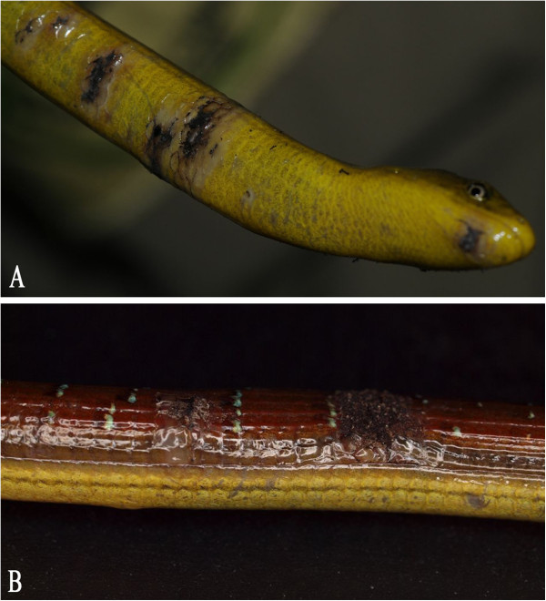
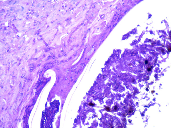
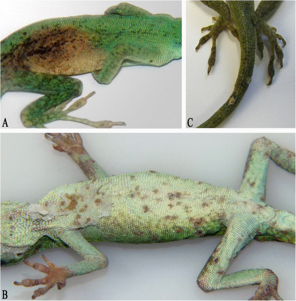
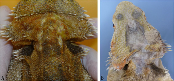
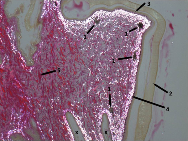
Ranaviral disease in amphibians has been studied intensely during the last decade, as associated mass-mortality events are considered to be a global threat to wild animal populations. Several studies have also included other susceptible ectothermic vertebrates (fish and reptiles), but only very few cases of ranavirus infections in lizards have been previously detected. In this study, we focused on clinically suspicious lizards and tested these animals for the presence of ranaviruses. Virological screening of samples from lizards with increased mortality and skin lesions over a course of four years led to the detection of ranaviral infections in seven different groups. Affected species were: brown anoles (Anolis sagrei), Asian glass lizards (Dopasia gracilis), green anoles (Anolis carolinensis), green iguanas (Iguana iguana), and a central bearded dragon (Pogona vitticeps). Purulent to ulcerative-necrotizing dermatitis and hyperkeratosis were diagnosed in pathological examinations. All animals tested positive for the presence of ranavirus by PCR and a part of the major capsid protein (MCP) gene of each virus was sequenced. Three different ranaviruses were isolated in cell culture. The analyzed portions of the MCP gene from each of the five different viruses detected were distinct from one another and were 98.4-100% identical to the corresponding portion of the frog virus 3 (FV3) genome. This is the first description of ranavirus infections in these five lizard species. The similarity in the pathological lesions observed in these different cases indicates that ranaviral infection may be an important differential diagnosis for skin lesions in lizards.
Figures
References
-
- Gray MJ, Miller DL, Hoverman JT. Ecology and pathology of amphibian ranaviruses. Dis Aquat Organ. 2009;87:243–266. - PubMed
Publication types
MeSH terms
Substances
LinkOut - more resources
Full Text Sources
Other Literature Sources
Miscellaneous
