. 2013 Sep 28:13:240.
doi: 10.1186/1472-6882-13-240.

Instrument-assisted cross fiber massage increases tissue perfusion and alters microvascular morphology in the vicinity of healing knee ligaments

Affiliations

Instrument-assisted cross fiber massage increases tissue perfusion and alters microvascular morphology in the vicinity of healing knee ligaments

M Terry Loghmani et al. BMC Complement Altern Med. .

Abstract

Background: Ligament injuries are common clinical problems for which there are few established interventions. Instrument-assisted cross fiber massage (IACFM) was recently shown to accelerate the restoration of biomechanical properties in injured rodent knee medial collateral ligaments (MCL). The current study aimed to investigate the influence of IACFM on regional perfusion and vascularity in the vicinity of healing rodent knee MCL injuries.

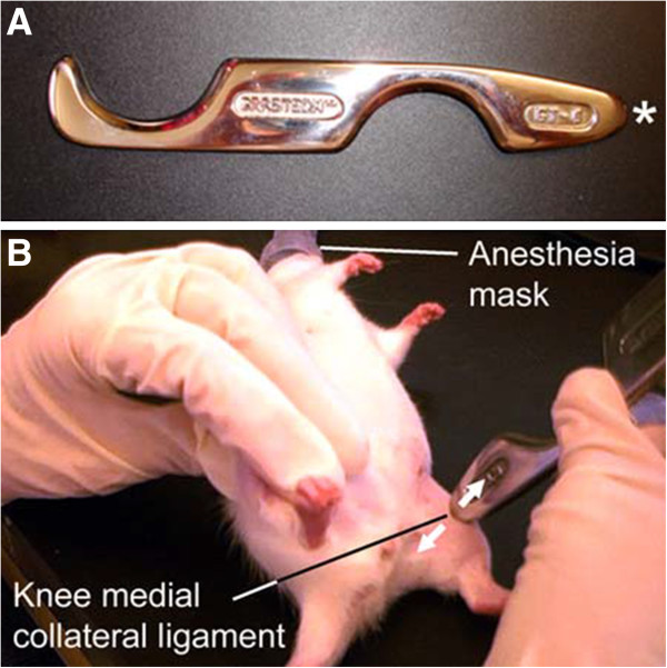
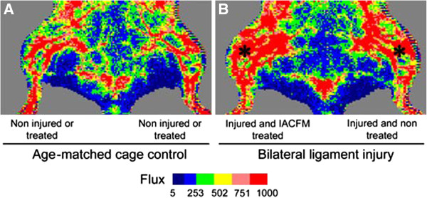
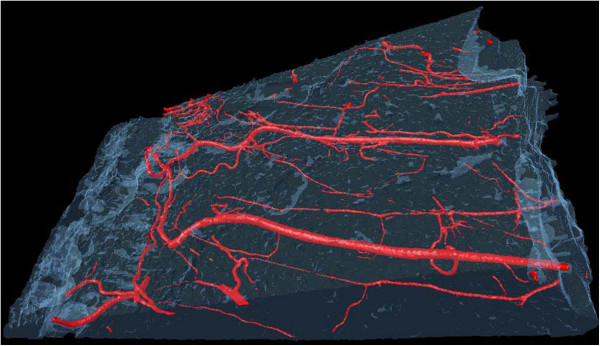
Methods: Bilateral knee MCL injuries were induced in female Sprague-Dawley rats. Commencing 1 week post-injury, 1 minute of IACFM was introduced unilaterally 3 times/week for 3 weeks. The contralateral injured MCL served as an internal control. Regional tissue perfusion was assessed in vivo throughout healing using laser Doppler imaging, whereas regional microvascular morphology was assessed ex vivo via micro-computed tomography of vessels filled with contrast.

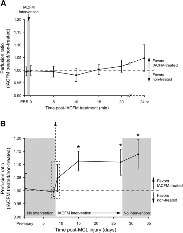
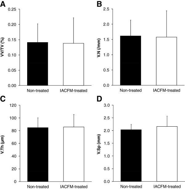
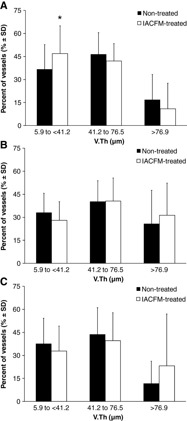
Results: IACFM had no effect on tissue perfusion when assessed immediately, or at 5, 10, 15 or 20 min following intervention (all p > 0.05). However, IACFM-treated hindlimbs had enhanced tissue perfusion when assessed 1 day following the 4th and 9th (last) treatment sessions (all p < 0.05). IACFM-treated hindlimbs also had greater perfusion when assessed 1 wk following the final treatment session (32 days post-injury) (p < 0.05). Subsequent investigation of microvascular morphology found IACFM to increase the proportion of arteriole-sized blood vessels (5.9 to <41.2 μm) in the tibial third of the ligament (p < 0.05).

Conclusions: These findings suggest IACFM alters regional perfusion and vascularity in the vicinity of healing rodent knee MCL injuries. This effect may contribute to the beneficial effect of IACFM observed on the recovery of knee ligament biomechanical properties following injury.


Figures

Figure 1
Figure 1
Instrument-assisted cross fiber massage (IACFM) intervention. A: The rigid Graston Technique® GT6 tool fabricated from stainless steel has a tapered tip (*) which permits treatment of small structures. B: IACFM of the rodent knee joint medial collateral ligament using the GT6 tool. Arrows indicate the direction of movement/force application perpendicular to the collagen substructure of the ligament.
Figure 2
Figure 2
Representative laser Doppler images for the left and right hindlimbs in A: aged-matched uninjured control animal and B: animal with bilateral knee MCL injuries (*) at four weeks post-injury. A: Tissue flux (perfusion) was equivalent in the right and left legs of age-matched control animals, animals prior to surgically-induced MCL injury, and animals immediately prior to the initial IACFM treatment (i.e. one week following MCL injury). B: Tissue flux (perfusion) was increased in the IACFM treated hindlimb at 4 weeks following injury and 1 week following the final IACFM treatment, compared to the contralateral non-treated injured hindlimb.
Figure 3
Figure 3
Effect of IACFM on regional tissue perfusion following knee MCL injury. A: IACFM had no immediate effect on tissue perfusion ratios, with ratios immediately prior (PRE) and after (0) the first IACFM treatment, at 5, 10, 15 and 20 minutes post-treatment, and at 24 hours post-treatment not differing significantly from 1 (i.e. perfusion in IACFM-treated = perfusion in non-treated). B: IACFM significantly increased tissue perfusion over time. In addition to having increased perfusion when assessed at 24 hours after the 4th and last (9th) treatment (15 and 26 days post-MCL injury, respectively), IACFM treated hindlimbs had significantly greater perfusion when assessed one week following the final treatment session (32 days post-MCL injury). Data represent perfusion ratios (IACFM treated/non treated) between IACFM treated and non-treated hindlimbs, with error bars indicating the 95% confidence interval. Ratios >1 indicate greater perfusion in IACFM-treated hindlimbs. *p < 0.05, as determined by single sample t-tests with a population mean of 1.
Figure 4
Figure 4
Representative micro-CT image of contrast agent perfused blood vessels within a MCL and its surrounding connective tissue. Microvasculature morphology parameters acquired from the whole tissue, and upper (femoral), middle and lower (tibial) thirds of the tissue volume were: vessel volume normalized to tissue volume (VV/TV; %), vessel number (V.N;/mm), vessel thickness or diameter (V.Th; μm), and vessel separation (V.Sp; mm). The MCL is not distinguishable within the tissue volume because of its equivalent radiopacity. Images from both IACFM-treated and non-treated hind limbs are not shown as they are visually indistinguishable.
Figure 5
Figure 5
Effect of IACFM on microvasculature morphometric parameters following knee MCL injury. There were no significant IACFM effects on A: vessel volume normalized to tissue volume [VV/TV], B: vessel number [V.N], C: vessel thickness or diameter [V.Th], or D: vessel number [V.N] (all p = 0.37 to 0.90). Bars represent mean ± SD.
Figure 6
Figure 6
Effect of IACFM on the frequency distribution of vessel thickness (V.Th). V.Th distribution in the A: tibial, B: middle, and C: femoral subregions of the injured knee MCL and adjacent connective tissue. IACFM-treated hindlimbs had a higher proportion of V.Th within the 5.9 to <41.2 μm range in the tibial subregion. No differences were found in the middle or femoral subregions. Bars represent mean ± SD. *indicates p < 0.02 compared to non-treated hindlimb.

References

    1. Fernandez WG, Yard EE, Comstock RD. Epidemiology of lower extremity injuries among U.S. high school athletes. Acad Emerg Med. 2007;14(7):641–645. - PubMed
    1. Konradsen L, Bech L, Ehrenbjerg M, Nickelsen T. Seven years follow-up after ankle inversion trauma. Scand J Med Sci Sports. 2002;12(3):129–135. doi: 10.1034/j.1600-0838.2002.02104.x. - DOI - PubMed
    1. Tyler TF, McHugh MP, Mirabella MR, Mullaney MJ, Nicholas SJ. Risk factors for noncontact ankle sprains in high school football players: the role of previous ankle sprains and body mass index. Am J Sports Med. 2006;34(3):471–475. - PubMed
    1. Valderrabano V, Hintermann B, Horisberger M, Fung TS. Ligamentous posttraumatic ankle osteoarthritis. Am J Sports Med. 2006;34(4):612–620. doi: 10.1177/0363546505281813. - DOI - PubMed
    1. van Rijn RM, van Os AG, Bernsen RM, Luijsterburg PA, Koes BW, Bierma-Zeinstra SM. What is the clinical course of acute ankle sprains? A systematic literature review. Am J Med. 2008;121(4):324–331. doi: 10.1016/j.amjmed.2007.11.018. - DOI - PubMed

Publication types