. 2013 Sep 28:14:95.
doi: 10.1186/1471-2156-14-95.

Mutation in MEOX1 gene causes a recessive Klippel-Feil syndrome subtype

Affiliations

Mutation in MEOX1 gene causes a recessive Klippel-Feil syndrome subtype

Fatih Bayrakli et al. BMC Genet. .

Abstract

Background: Klippel-Feil syndrome (KFS) is characterized by the developmental failure of the cervical spine and has two dominantly inherited subtypes. Affected individuals who are the children of a consanguineous marriage are extremely rare in the medical literature, but the gene responsible for this recessive trait subtype of KFS has recently been reported.

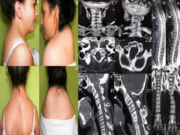
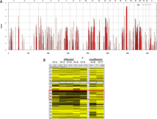
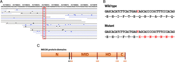
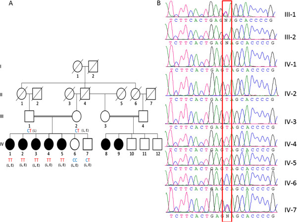
Results: We identified a family with the KFS phenotype in which their parents have a consanguineous marriage. Radiological examinations revealed that they carry fusion defects and numerical abnormalities in the cervical spine, scoliosis, malformations of the cranial base, and Sprengel's deformity. We applied whole genome linkage and whole-exome sequencing analysis to identify the chromosomal locus and gene mutated in this family. Whole genome linkage analysis revealed a significant linkage to chromosome 17q12-q33 with a LOD score of 4.2. Exome sequencing identified the G > A p.Q84X mutation in the MEOX1 gene, which is segregated based on pedigree status. Homozygous MEOX1 mutations have reportedly caused a similar phenotype in knockout mice.

Conclusions: Here, we report a truncating mutation in the MEOX1 gene in a KFS family with an autosomal recessive trait. Together with another recently reported study and the knockout mouse model, our results suggest that mutations in MEOX1 cause a recessive KFS phenotype in humans.


Figures

Figure 1
Figure 1
Patient photographs of family members IV-2 and IV-3 show KFS phenotypes. Photographs of patients from the side (A and B) and behind (C and D) show short neck, low posterior hairline, and elevated scapula (Sprengel’s deformity). 3D cervical spine skull base CT images show omovertebral bone causing Sprengel’s deformity, posterior neural arch fusion defects, and fusions between vertebrae (E and F). Whole spine sagittal section T2-weighted MR images show cervical vertebrae count anomaly (G and H). Axial skull base CT images show malformation of the foramen magnum (I and J). Sagittal reconstruction of cervical and skull base CT images show abnormal bony formation emerging from posterior structures as well as fusion between posterior structures of some successive vertebrae, occipitalization of atlas and dens, and corpus anomalies (K and L). Scoliosis is evident on coronal T2-weighted whole spine MR images. Fusion defects are seen in midportions of vertebra corpuses (M and N).
Figure 2
Figure 2
Genome-wide linkage analysis with and homozygosity mapping 250 K Nsp I Affymetrix Gene Chips. (A) the locus on chromosome 17 was the only one to give the theoretical maximum for this pedigree. cM, centimorgan; LOD, logarithm of odds. (B) homozygosity mapping of KFS family via autoSNPa. Each column represents the homozygosity profile of one individual, and each row represents the call of an individual SNP on chromosome 17 (yellow, heterozygous; black, homozygous). Note the large run of homozygosity that is exclusively shared by the affected members (boxed in red).
Figure 3
Figure 3
MEOX1 mutation analysis. (A) G → A nucleotide change (red box) in MEOX1 is identified by exome sequencing. (B) This nucleotide change causes the formation of a stop codon and a truncated protein. (C) The mutation is located at the end of the N terminal domain (N: N terminal domain, MID: middle domain, HD: home domain, C: C terminal domain; black vertical line shows mutation location).
Figure 4
Figure 4
KFS family pedigree. (A) Affected sibs are identified by filled symbols. Diagonal lines indicate deceased family members. Circles represent female and squares represent male family members. The affected genotype is TT, healthy individuals are CT and CC genotypes. “L” represents patients used in whole genome linkage analysis; “E” represents those used in whole-exome sequencing. (B) Sanger sequencing confirms the altered base in family members (red box).

References

    1. Tracy MR, Dormans JP, Kusumi K. Klippel-Feil syndrome: clinical features and current understanding of etiology. Clin Orthop Relat Res. 2004;424:183–190. - PubMed
    1. Kaplan KM, Spivak JM, Bendo JA. Embryology of the spine and associated congenital abnormalities. Spine J. 2005;5(5):564–576. doi: 10.1016/j.spinee.2004.10.044. - DOI - PubMed
    1. Harvey EJ, Bernstein M, Desy NM, Saran N, Ouellet JA. Sprengel deformity: pathogenesis and management. J Am Acad Orthop Surg. 2012;20(3):177–186. doi: 10.5435/JAAOS-20-03-177. - DOI - PubMed
    1. Saga Y. The mechanism of somite formation in mice. Curr Opin Genet Dev. 2012;22(4):331–338. doi: 10.1016/j.gde.2012.05.004. - DOI - PubMed
    1. Skuntz S, Mankoo B, Nguyen MT, Hustert E, Nakayama A, Tournier-Lasserve E, Wright CV, Pachnis V, Bharti K, Arnheiter H. Lack of the mesodermal homeodomain protein MEOX1 disrupts sclerotome polarity and leads to a remodeling of the cranio-cervical joints of the axial skeleton. Dev Biol. 2009;332(2):383–395. doi: 10.1016/j.ydbio.2009.06.006. - DOI - PMC - PubMed

Publication types

MeSH terms

LinkOut - more resources