. 2014 Feb 15;23(4):406-20.
doi: 10.1089/scd.2013.0412. Epub 2013 Nov 7.

Genome wide profiling of dopaminergic neurons derived from human embryonic and induced pluripotent stem cells

Affiliations

Genome wide profiling of dopaminergic neurons derived from human embryonic and induced pluripotent stem cells

Olga Momčilović et al. Stem Cells Dev. .

Abstract

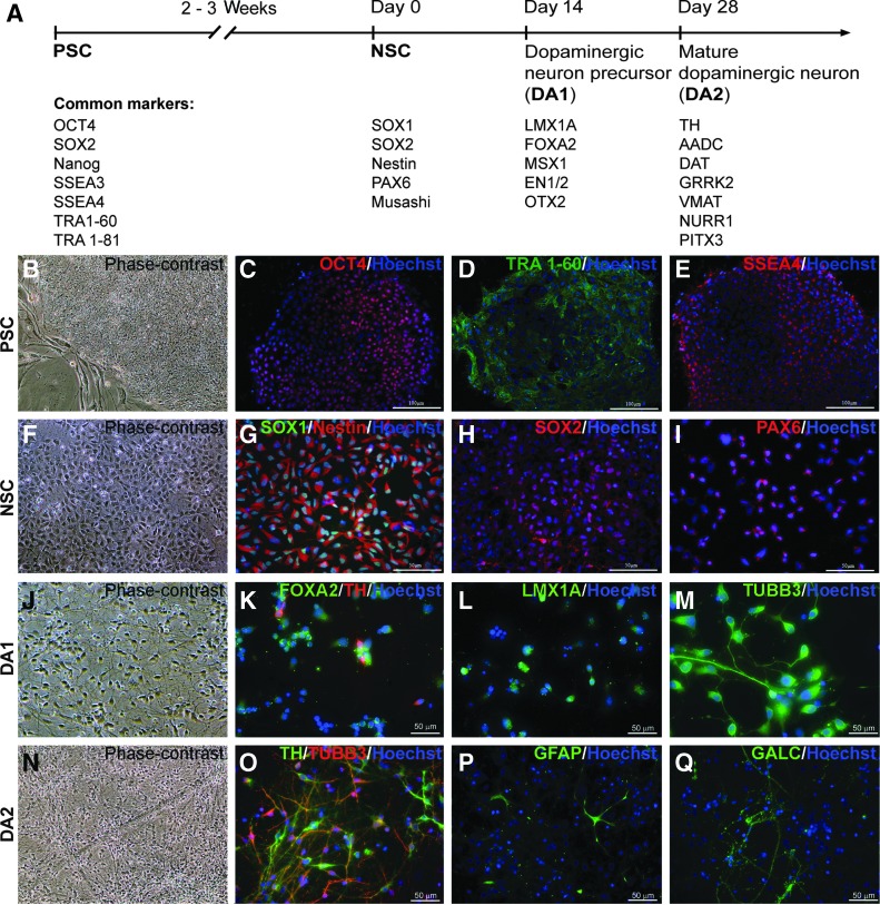
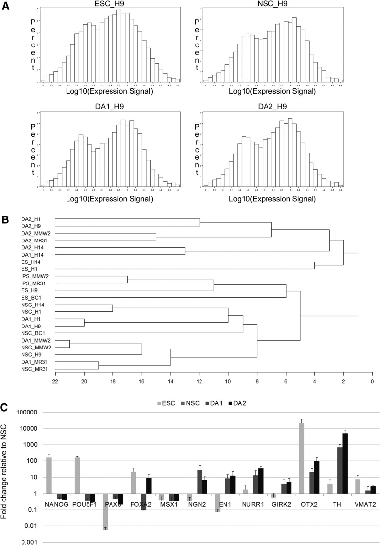
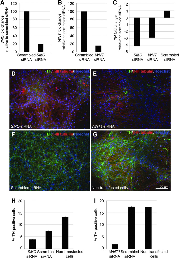
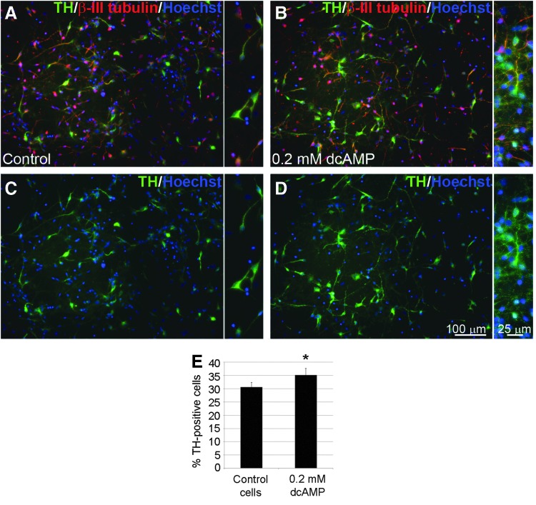
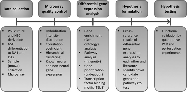
Recent advances in human embryonic stem cell (ESC) and induced pluripotent stem cell (iPSC) biology enable generation of dopaminergic neurons for potential therapy and drug screening. However, our current understanding of molecular and cellular signaling that controls human dopaminergic development and function is limited. Here, we report on a whole genome analysis of gene expression during dopaminergic differentiation of human ESC/iPSC using Illumina bead microarrays. We generated a transcriptome data set containing the expression levels of 28,688 unique transcripts by profiling five lines (three ESC and two iPSC lines) at four stages of differentiation: (1) undifferentiated ESC/iPSC, (2) neural stem cells, (3) dopaminergic precursors, and (4) dopaminergic neurons. This data set provides comprehensive information about genes expressed at each stage of differentiation. Our data indicate that distinct pathways are activated during neural and dopaminergic neuronal differentiation. For example, WNT, sonic hedgehog (SHH), and cAMP signaling pathways were found over-represented in dopaminergic populations by gene enrichment and pathway analysis, and their role was confirmed by perturbation analyses using RNAi (small interfering RNA of SHH and WNT) or small molecule [dibutyryl cyclic AMP (dcAMP)]. In summary, whole genome profiling of dopaminergic differentiation enables systematic analysis of genes/pathways, networks, and cellular/molecular processes that control cell fate decisions. Such analyses will serve as the foundation for better understanding of dopaminergic development, function, and development of future stem cell-based therapies.


Figures

<b>FIG. 1.</b>
FIG. 1.
Stage-specific dopaminergic differentiation protocol. (A) Schematic depiction of the differentiation protocol with the list of representative markers at the each stage. (B–E) Phase contrast (B) and immunocytochemistry images in pluripotent stem cells (PSCs) with antibodies against OCT4 (C), TRA1–60 (D), and SSEA4 (E). (F–I) Phase contrast (F) and immunocytochemistry images in neural stem cells (NSC) with antibodies against SOX1/Nestin (G), SOX2 (H), and PAX6 (I). (J–M) Phase contrast (J) and immunocytochemistry images in dopaminergic precursors (DA1) with antibodies against FOXA2/TH (K), LMX1A (L), and β-III-tubulin (M). (N–Q) Phase contrast (N) and immunocytochemistry images in mature dopaminergic population (DA2) for neuronal/dopaminergic markers β-III-tubulin/TH (O), astrocyte marker GFAP (P), and oligodendrocyte marker GALC (Q). Blue—DNA. Scale bar as marked.
<b>FIG. 2.</b>
FIG. 2.
Quality control analysis of microarray data. (A) Histogram representation of percentage of genes with different expression intensities for four H9 samples—embryonic stem cell (ESC), NSC, DA1, and DA2. (B) Unsupervised hierarchical clustering of gene expression data for all 20 samples from five PSC lines at four differentiation stages. (C) Quantitative polymerase chain reaction (qPCR) validation of microarray data. GAPDH and TBP served as endogenous standards, and data were normalized against NSC sample. Note the use of logarithmic scale on the y-axis (ie, the x-axis crosses the y-axis at 1, which is also the relative gene expression in NSC).
<b>FIG. 3.</b>
FIG. 3.
The effect of repression of sonic hedgehog (SHH) and WNT1 signaling pathways on dopaminergic differentiation. (A–C) qPCR analysis of SMO (A), WNT1 (B), and TH (C) gene expression following SMO (A), WNT1 (B), SMO, or WNT1 small interfering RNA (siRNA) treatment. (D–G) Representative TH (green) and β-III-tubulin (red) immunocytochemistry images following SMO (D), WNT1 (E), scrambled siRNA treatment (F), or in nontransfected cells (G). Scale bar as marked. (H, I) Quantification of TH-positive cells following SMO siRNA (H) or WNT1 siRNA (I) treatment.
<b>FIG. 4.</b>
FIG. 4.
Dibutyryl cyclic AMP (dcAMP) promotes dopaminergic differentiation. (A–D) Representative TH (green) and β-III-tubulin (red) immunocytochemistry images in control (A, B) and 0.2 mM dcAMP-treated (C, D) cells; inserts—higher magnification. Scale bar as marked. (E) Quantification of TH-positive cells in control and dcAMP-treated samples. The asterisk denotes 0.01<P value <0.05.
<b>FIG. 5.</b>
FIG. 5.
The process of microarray data analysis. After mRNA isolation and data collection, we performed a set of quality control steps to ensure the quality of data. In the following steps differentially expressed genes were analyzed and such results were cross-referenced with literature to form hypotheses and validate obtained results.

References

    1. Takahashi K, Tanabe K, Ohnuki M, Narita M, Ichisaka T, Tomoda K. and Yamanaka S. (2007). Induction of pluripotent stem cells from adult human fibroblasts by defined factors. Cell 131:861–872 - PubMed
    1. Yu J, Vodyanik MA, Smuga-Otto K, Antosiewicz-Bourget J, Frane JL, Tian S, Nie J, Jonsdottir GA, Ruotti V, et al. (2007). Induced pluripotent stem cell lines derived from human somatic cells. Science 318:1917–1920 - PubMed
    1. Zeng X, Cai J, Chen J, Luo Y, You ZB, Fotter E, Wang Y, Harvey B, Miura T, et al. (2004). Dopaminergic differentiation of human embryonic stem cells. Stem Cells 22:925–940 - PubMed
    1. Swistowski A, Peng J, Han Y, Swistowska AM, Rao MS. and Zeng X. (2009). Xeno-free defined conditions for culture of human embryonic stem cells, neural stem cells and dopaminergic neurons derived from them. PLoS One 4:e6233. - PMC - PubMed
    1. Kriks S, Shim JW, Piao J, Ganat YM, Wakeman DR, Xie Z, Carrillo-Reid L, Auyeung G, Antonacci C, et al. (2011). Dopamine neurons derived from human ES cells efficiently engraft in animal models of Parkinson's disease. Nature 480:547–551 - PMC - PubMed

Publication types

Substances

LinkOut - more resources