

Altered cerebral perfusion in executive, affective, and motor networks during adolescent depression
- PMID: 24074474
- PMCID: PMC3825460
- DOI: 10.1016/j.jaac.2013.07.008
Altered cerebral perfusion in executive, affective, and motor networks during adolescent depression
Abstract
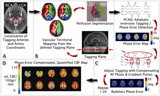
Objective: Although substantial literature has reported regional cerebral blood flow (rCBF) abnormalities in adults with depression, these studies commonly necessitated the injection of radioisotopes into subjects. The recent development of arterial spin labeling (ASL), however, allows noninvasive measurements of rCBF. Currently, no published ASL studies have examined cerebral perfusion in adolescents with depression. Thus, the aim of the present study was to examine baseline cerebral perfusion in adolescent depression using a newly developed ASL technique: pseudocontinuous arterial spin labeling (PCASL).
Method: A total of 25 medication-naive adolescents (13-17 years of age) diagnosed with major depressive disorder (MDD) and 26 well-matched control subjects underwent functional magnetic resonance imaging. Baseline rCBF was measured via a novel PCASL method that optimizes tagging efficiency.
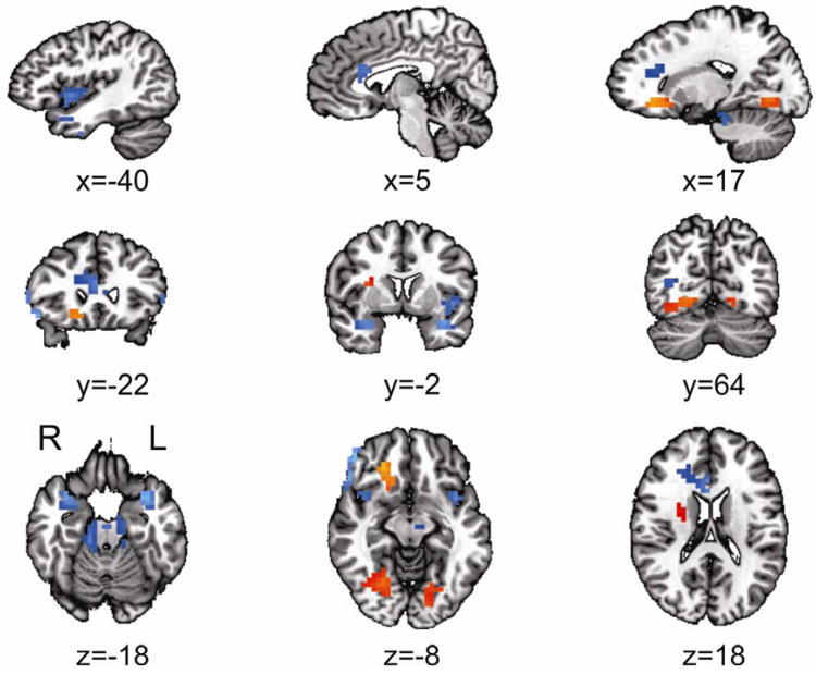
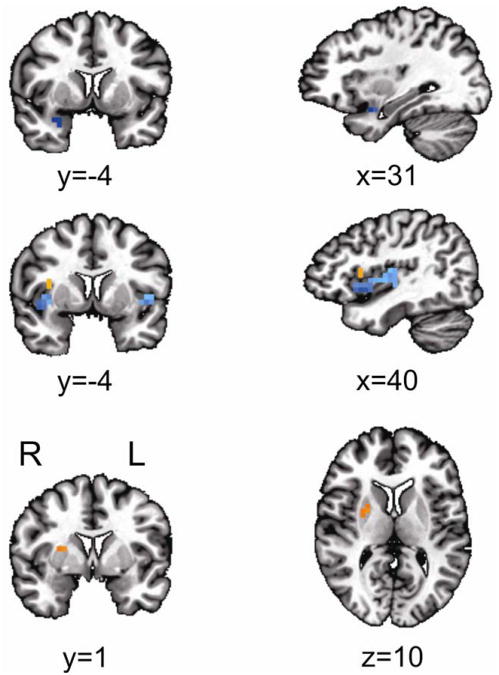
Results: Voxel-based whole brain analyses revealed significant frontal, limbic, paralimbic, and cingulate hypoperfusion in the group with depression (p < .05, corrected). Hyperperfusion was also observed within the subcallosal cingulate, putamen, and fusiform gyrus (p < .05, corrected). Similarly, region-of-interest analyses revealed amygdalar and insular hypoperfusion in the group with depression, as well as hyperperfusion in the putamen and superior insula (p < .05, corrected).
Conclusions: Adolescents with depression and healthy adolescents appear to differ on rCBF in executive, affective, and motor networks. Dysfunction in these regions may contribute to the cognitive, emotional, and psychomotor symptoms commonly present in adolescent depression. These findings point to possible biomarkers for adolescent depression that could inform early interventions and treatments, and establishes a methodology for using PCASL to noninvasively measure rCBF in clinical and healthy adolescent populations.
Keywords: cerebral blood flow; functional magnetic resonance imaging (fMRI); major depressive disorder (MDD); pseudocontinuous arterial spin labeling (PCASL).
Copyright © 2013 American Academy of Child and Adolescent Psychiatry. Published by Elsevier Inc. All rights reserved.
Figures
Similar articles
-
Characteristic distributions of regional cerebral blood flow changes in major depressive disorder patients: a pseudo-continuous arterial spin labeling (pCASL) study.J Affect Disord. 2014 Aug;165:59-63. doi: 10.1016/j.jad.2014.04.032. Epub 2014 Apr 21. J Affect Disord. 2014. PMID: 24882178
-
Regional cerebral blood flow in major depression treated with electroconvulsive therapy: an arterial spin labeling magnetic resonance study.Neurocase. 2022 Apr;28(2):246-250. doi: 10.1080/13554794.2022.2044861. Epub 2022 Feb 27. Neurocase. 2022. PMID: 35225161
-
Arterial spin labeling reveals relationships between resting cerebral perfusion and motor learning in Parkinson's disease.Brain Imaging Behav. 2019 Jun;13(3):577-587. doi: 10.1007/s11682-018-9877-1. Brain Imaging Behav. 2019. PMID: 29744796 Free PMC article.
-
Characterization of brain blood flow and the amplitude of low-frequency fluctuations in major depressive disorder: A multimodal meta-analysis.J Affect Disord. 2017 Mar 1;210:303-311. doi: 10.1016/j.jad.2016.12.032. Epub 2016 Dec 23. J Affect Disord. 2017. PMID: 28068619 Review.
-
Electroconvulsive therapy-induced neuroimaging alterations measured by cerebral blood flow in adolescents with major depressive disorder.J Affect Disord. 2023 Apr 14;327:385-390. doi: 10.1016/j.jad.2023.02.027. Epub 2023 Feb 8. J Affect Disord. 2023. PMID: 36758871 Review.
Cited by
-
Effects of the antidepressant medication duloxetine on brain metabolites in persistent depressive disorder: A randomized, controlled trial.PLoS One. 2019 Jul 19;14(7):e0219679. doi: 10.1371/journal.pone.0219679. eCollection 2019. PLoS One. 2019. PMID: 31323045 Free PMC article. Clinical Trial.
-
Associations between depression and anxiety symptoms and retinal vessel caliber in adolescents and young adults.Psychosom Med. 2014 Nov-Dec;76(9):732-8. doi: 10.1097/PSY.0000000000000117. Psychosom Med. 2014. PMID: 25373892 Free PMC article.
-
A neuroradiologist's guide to arterial spin labeling MRI in clinical practice.Neuroradiology. 2015 Dec;57(12):1181-202. doi: 10.1007/s00234-015-1571-z. Epub 2015 Sep 9. Neuroradiology. 2015. PMID: 26351201 Free PMC article. Review.
-
The Cerebral Blood Flow Biomedical Informatics Research Network (CBFBIRN) data repository.Neuroimage. 2016 Jan 1;124(Pt B):1202-1207. doi: 10.1016/j.neuroimage.2015.05.059. Epub 2015 May 30. Neuroimage. 2016. PMID: 26032887 Free PMC article.
-
Causal connectivity alterations of cortical-subcortical circuit anchored on reduced hemodynamic response brain regions in first-episode drug-naïve major depressive disorder.Sci Rep. 2016 Feb 25;6:21861. doi: 10.1038/srep21861. Sci Rep. 2016. PMID: 26911651 Free PMC article.
References
-
- The World Health Organization. The global burden of disease: 2004 update, Table 9: Estimated prevalence of moderate and severe disability for leading disabling conditions by age, for high-income and low- and middle-income countries, 2004. 2008 http://www.who.int/healthinfo/global_burden_disease/GBD_report2004update....
-
- Kessler RC, Berglund P, Demler O, et al. The epidemiology of major depressive disorder: results from the National Comorbidity Survey Replication (NCS-R) JAMA : the journal of the American Medical Association. 2003 Jun 18;289(23):3095–3105. - PubMed
-
- Kessler RC, Angermeyer M, Anthony JC, et al. Lifetime prevalence and age-of-onset distributions of mental disorders in the World Health Organization's World Mental Health Survey Initiative. World psychiatry : official journal of the World Psychiatric Association (WPA) 2007 Oct;6(3):168–176. - PMC - PubMed
-
- Pine DS, Cohen E, Cohen P, Brook J. Adolescent depressive symptoms as predictors of adult depression: moodiness or mood disorder? The American journal of psychiatry. 1999 Jan;156(1):133–135. - PubMed
-
- Davidson RJ, Pizzagalli D, Nitschke JB, Putnam K. Depression: perspectives from affective neuroscience. Annual review of psychology. 2002;53:545–574. - PubMed
Publication types
MeSH terms
Substances
Grants and funding
LinkOut - more resources
Full Text Sources
Other Literature Sources
Medical
