. 2013 Sep 26;155(1):228-41.
doi: 10.1016/j.cell.2013.08.042.

Maternal and offspring pools of osteocalcin influence brain development and functions

Affiliations

Maternal and offspring pools of osteocalcin influence brain development and functions

Franck Oury et al. Cell. .

Abstract

The powerful regulation of bone mass exerted by the brain suggests the existence of bone-derived signals modulating this regulation or other functions of the brain. We show here that the osteoblast-derived hormone osteocalcin crosses the blood-brain barrier, binds to neurons of the brainstem, midbrain, and hippocampus, enhances the synthesis of monoamine neurotransmitters, inhibits GABA synthesis, prevents anxiety and depression, and favors learning and memory independently of its metabolic functions. In addition to these postnatal functions, maternal osteocalcin crosses the placenta during pregnancy and prevents neuronal apoptosis before embryos synthesize this hormone. As a result, the severity of the neuroanatomical defects and learning and memory deficits of Osteocalcin(-/-) mice is determined by the maternal genotype, and delivering osteocalcin to pregnant Osteocalcin(-/-) mothers rescues these abnormalities in their Osteocalcin(-/-) progeny. This study reveals that the skeleton via osteocalcin influences cognition and contributes to the maternal influence on fetal brain development.


Figures

Figure 1
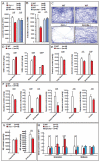
Figure 1. Osteocalcin affects the biosynthesis of neurotransmitters
Measurements of (A) total (XTOT) and (B) ambulatory activity (AMBX), in Ocn−/− and Gprc6a−/− mice, during 12h light and dark phases over 3 days. Mutant mice were compared to their WT littermates. (C) Video tracking of an open field paradigm test performed in Ocn−/−, Gprc6a−/− and WT littermate mice. (D) GABA, (E) serotonin, (F) dopamine, and (G) norepinephrine contents in various parts of the brains of Ocn−/− and WTmice. (H) Expression (qPCR) of Tryptophan hydroxylase-2 (Tph2), Glutamate decarboxylase-1 and -2 (Gad1), (Gad2) and Tyrosine hydroxylase (Th) in the brainstem and midbrain of Ocn−/−, Gprc6a−/− and control mice. Error bars represent SEM. The statistical test on the top of each graph represents the Student’s t test, p<0.05 is significant.
Figure 2
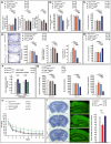
Figure 2. Osteocalcin affects several behaviors
(A) Light and Dark test (L/DT): The latency (Sec) to enter the lit compartment, number of transitions between compartments and time spent in the lit compartment were measured. (B) Elevated Plus Maze test (EPMT): Number of entries and time spent (Sec) in the open arms were scored. (C) Open field test (OFT): Total distance (cm), % of the distance traveled in the center versus periphery, % of the time spent in the center versus periphery and number of rearing events were measured. The video tracking of each group of mice is represented on the right panel. (D-E) Representation of the time spent (Sec) immobile during the (D) Tail Suspension test and (E) the Forced Swim test. Both tests assess depression-like behavior. (F-G) Morris Water Maze test performed over 12 days. The graphic shows the time (seconds) needed for each group of mice to localize a submerged platform in the swimming area. The video tracking on the left panel represents the standards obtained for each group analyzed. Error bars represent SEM. The statistical test on the top of each graph represents the Student’s t test, (a) means P value of < 0.05 versus WT, p<0.05 is significant.
Figure 3
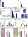
Figure 3. Osteocalcin binds to neurons in the brain
(A) Measurement of osteocalcin in bone, serum, cortex, midbrain, hypothalamus and brainstem of Ocn−/− mice receiving either PBS (first column), uncarboxylated (second column), or carboxylated (third column) osteocalcin s.c. during 7 days. (B) Binding of GST-biotin (1.5μg/ml) (panel 1) and biotinylated uncarboxylated osteocalcin (300ng/ml) (panels2-4) to the dorsal (DR) (first row) and median (MR) (second row) raphe nuclei of the brainstem (identified by anti-5-HT immunofluorescence), to the ventral tegmental area (VTA) (third row) of the midbrain (identified by anti-TH immunofluorescence) and to the CA3 region (fourth row) of the hippocampus (identified anatomically). Panels 5: competition with unlabeled osteocalcin (1000-fold excess). Scale bars = 100μm (C) Expression of Tph2 and Gad1 in brainstem, of Th in midbrain explants from WT and Gprc6a−/− mice, treated with 3ng/ml osteocalcin or vehicle. (D) Gene expression in mouse primary hindbrain neurons treated with osteocalcin (10ng/ml) or vehicle. (E) Calcium flux of hindbrain neurons responsive (n=7) and unresponsive (n=5) to osteocalcin (30ng/ml). (F-G) Extracellular current recordings of (F) neurons of the dorsal raphe nucleus and (G) GABAergic interneurons of the brainstem treated with osteocalcin (10ng/ml). Error bars represent SEM. The statistical test on the top of each graph represents the Student’s t test, p<0.05 is significant.
Figure 4
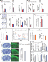
Figure 4. Osteocalcin signaling in the brain prevents anxiety and depression
(A) Light and Dark, (B) Elevated plus maze, (C) Open field, (D) Forced Swim and (E) Tail suspension tests performed in WT (n=7) and Ocn−/− mice infused with vehicle (n=7) or osteocalcin (10ng/hour) (n=7). (F) Expression (qPCR) of Tph2, Gad1 and Gad2 in brainstem, and Th in midbrain of WT (n=6) and Ocn−/− mice infused with vehicle (n=6) or uncarboxylated mouse osteocalcin (10ng/hour) (n=7). (G) Gene expression analysis (qPCR) in bone and in the cortex, striatum, hippocampus, hypothalamus, midbrain, brainstem and cerebellum in WT and Ocn−/− mice. (H) In situ hybridization of Ocn in WT and Ocn−/− adult brain (sagittal sections are represented) (Scale bars = 1mm) and in WT bone as a control (Scale bars = 0.5mm). Error bars represent SEM. The statistical test on the top of each graph represents the Student’s t test, p<0.05 is significant.
Figure 5
Figure 5. Osteocalcin regulates brain functions post-natally
(A) Light and dark, (B) Elevated plus maze, (C) Forced Swim, (D) Open field, (E) Tail suspension and (H) Morris Water maze tests performed in a cohort of Ocnflox/flox , □1Col1-CreERT2 OcnosbERT2 and mice. (F) Expression (qPCR) of Gad1 and Gad2 in brainstem and striatum of Ocnflox/flox, □1Col1-CreERT2 and OcnosbERT2 mice. (G) HPLC analysis of serotonin, dopamine and norepinephrine contents in brainstem, midbrain and striatum of Ocnflox/flox, □1Col1-CreERT2 and OcnosbERT2 mice. (I) Cresyl violet staining and NeuN immunofluorescence of control Oc□̅ and OcnosbERT2 hippocampi. The dentate gyrus area (% versus WT) is represented in the right panel. In each panel (A-F and H), “Total Control” means □1Col1-CreERT2 and Ocnflox/flox mice combined. Scale bars = 0.5mm. Error bars represent SEM. The statistical test on the top of each graph represents the Student’s t test, p<0.05 is significant.
Figure 6
Figure 6. Maternal osteocalcin favors fetal neurogenesis
(A) Expression of Ocn (qPCR) in bone, brain and placenta of WT and Ocn−/− newborn (P0) and embryos (E13.5 to E18.5). (B) Osteocalcin circulating levels in WT or Ocn−/− newborns (P0) and embryos (E13.5 to E18.5). (C) Ex vivo dual perfusion system that monitors the transport of osteocalcin across the placenta. Uncarboxylated mouse osteocalcin (300 ng/ml) was injected through the uterine artery in placentas obtained from WT mice at E14.5, 15.5, and 18.5 of pregnancy. Osteocalcin in fetal eluates is represented as % of maternal input. (D) Circulating levels of osteocalcin in WT embryos originating from WT or Ocn+/− mothers, of Ocn+/− embryos originating from Ocn+/− or Ocn−/− mothers and of Ocn−/− embryos originating from Ocn+/− or Ocn−/− mothers. Measurements were performed at E16.5 and E18.5. (E) Cresyl violet stain of lateral ventricles of hippocampi of E18.5 WT embryos originating from WT mothers and Ocn−/− embryos originating from Ocn+/− or Ocn−/− mothers. The measurements of the lateral ventricle area over brain area are represented below the images (in %). (Scale bars = 0.5mm). (F) Number of apoptotic cells (stained by TUNNEL assay) in hippocampi of E18.5 WT embryos carried by WT mothers and Ocn−/− embryos carried by Ocn+/− or Ocn−/− mothers. (G-H) Contextual fear conditioning (CFC) (G) and novel object recognition (NOR) (H) performed in WT and Ocn−/− mice born from Ocn−/− or Ocn+/− mothers (n = 7-18 per group). In the CFC, Ocn−/− mice born from Ocn−/− mothers mice exhibited significantly less context-elicited freezing than WT mice in context A and A’. In the NOR, there was a significant increase in the exploratory period in Ocn−/− mice born from Ocn−/− mothers compared to Ocn−/− mice born from Ocn+/− mothers or WT mice when a novel object was introduced. (I-J) BrdU and DCX Immunohistochemistry showing a significantly lower number of BrdU+ (I) and DCX+ (J) cells in the dentate gyrus (DG) of WT and Ocn−/− mice born from Ocn−/− or Ocn+/− mothers. This decrease was even more pronounce in the ventral region of the DG. (Scale bars = 0.2mm) For (A-F and I-J), the statistical test on the top of each graph represents the Student’s t test, p<0.05 is significant. For (G-H), the statistical test on the top of each graph represents an ANOVA. Significant ANOVAs were follwed up with Fisher’s PLSD tests where appropriate. (*) P value < 0.05, (**) P value < 0.01, (***) P value < 0.001.
Figure 7
Figure 7. Maternal osteocalcin determines spatial learning and memory in adult offspring
(A) Light and Dark, (B) Elevated plus maze, (C) Open field, (D) Forced Swim, (E) Tail suspension and (F) Morris Water maze tests performed in 3 month-old Ocn−/− mice born from Ocn−/− mothers injected once a day with vehicle or osteocalcin (240ng/day) during pregnancy compared to WT mice. (G) Surface of the lateral ventricle over brain area (%) of E18.5 hippocampi coronal sections of WT embryos originating from WT mothers and Ocn−/− embryos originating from osteocalcin-injected Ocn−/− mothers. (H) Number of apoptotic cells (stained by TUNNEL assay) of E18.5 hippocampi coronal sections of WT embryos originating from WT mothers and Ocn−/− embryos originating from Ocn−/− mothers injected with osteocalcin (240ng/day). (I) Cresyl violet, NeuN immunofluorescence and dentate gyrus area (% versus WT) of WT and Ocn−/− originating from osteocalcin-injected Ocn−/−. Scale bars = 0.5mm. (J-K) HPLC measurements of serotonin (J), norepinephrine (NE) (J) content in the brainstem; dopamine content in the striatum (J), and GABA content in the brainstem, midbrain and striatum (K) of WT and Ocn−/− originating from osteocalcin-injected Ocn−/− mothers. Error bars represent SEM. The statistical test on the top of each graph represents the Student’s t test, p<0.05 is significant. For (F) (a) means P value of < 0.05 versus WT, (b) mean P value of < 0.05 versus Ocn−/− originating from Ocn−/− mothers injected, p<0.05 is significant.

Comment in

References

    1. Banks WA, Kastin AJ, Huang W, Jaspan JB, Maness LM. Leptin enters the brain by a saturable system independent of insulin. Peptides. 1996;17:305–311. - PubMed
    1. Broad KD, Keverne EB. Placental protection of the fetal brain during short-term food deprivation. Proceedings of the National Academy of Sciences of the United States of America. 2011;108:15237–15241. - PMC - PubMed
    1. Challis JR. Endocrine disorders in pregnancy: Stress responses in children after maternal glucocorticoids. Nature reviews Endocrinology. 2012;8:629–630. - PubMed
    1. Crawley JN. Exploratory behavior models of anxiety in mice. Neuroscience and biobehavioral reviews. 1985;9:37–44. - PubMed
    1. Cryan JF, Mombereau C, Vassout A. The tail suspension test as a model for assessing antidepressant activity: review of pharmacological and genetic studies in mice. Neuroscience and biobehavioral reviews. 2005;29:571–625. - PubMed

Publication types