. 2013 Oct 14;27(1):103-12.
doi: 10.1016/j.devcel.2013.08.021. Epub 2013 Sep 26.

Deuterosome-mediated centriole biogenesis

Affiliations

Deuterosome-mediated centriole biogenesis

Deborah A Klos Dehring et al. Dev Cell. .

Abstract

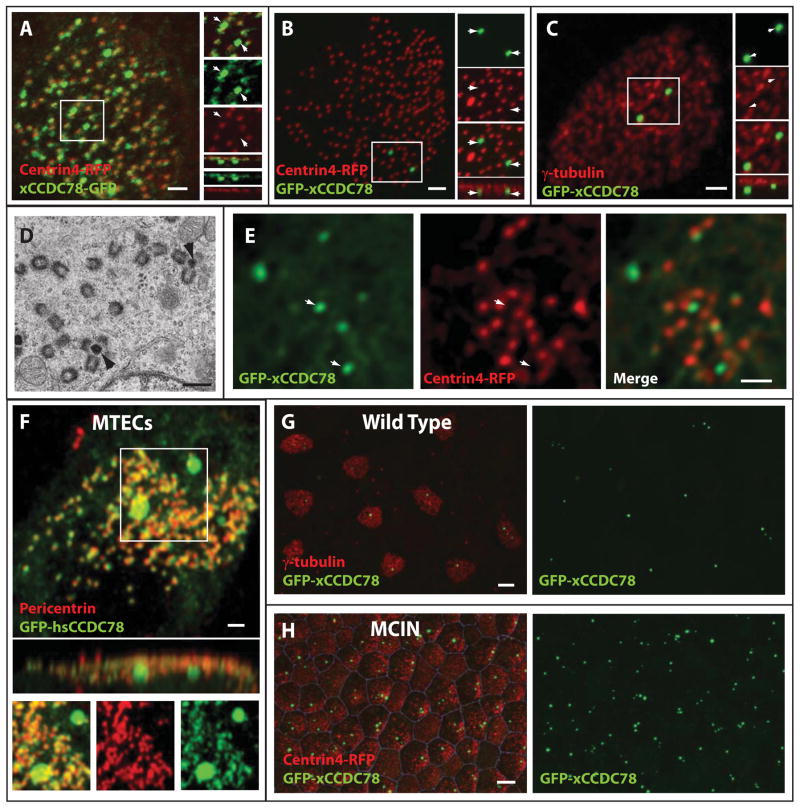
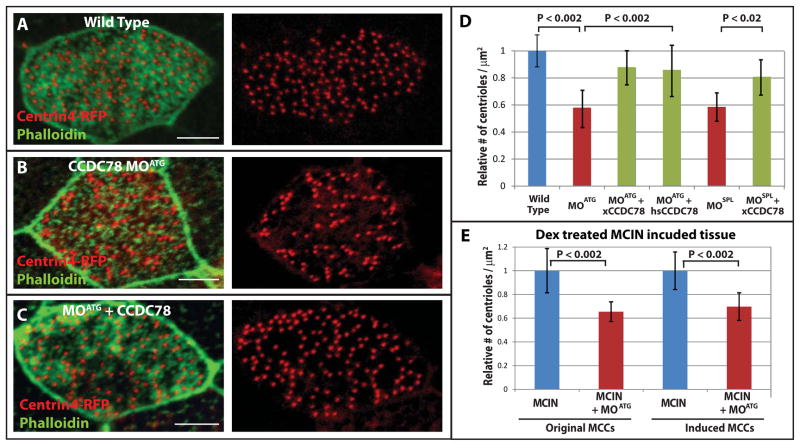
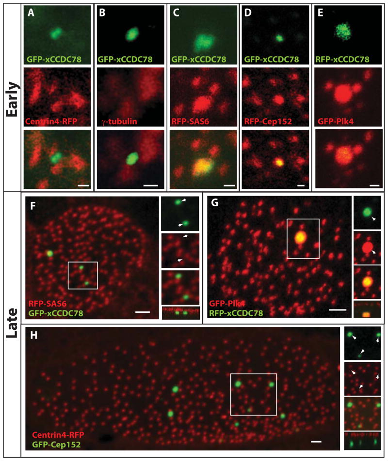
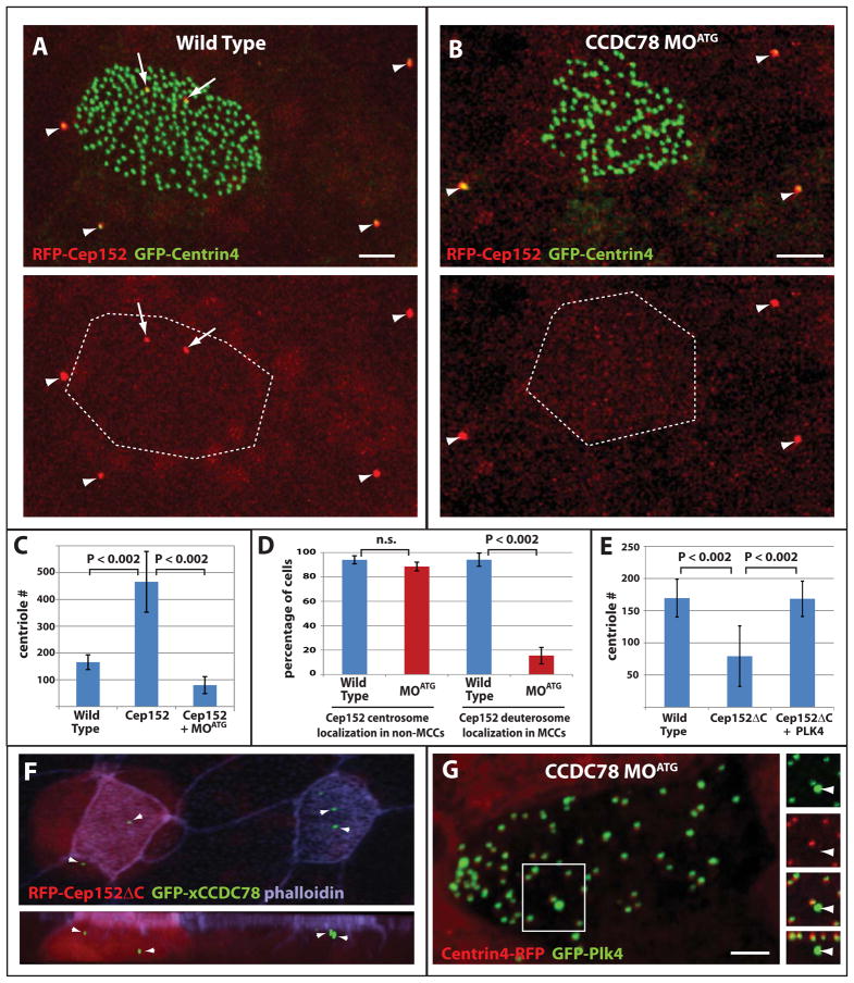
The ability of cells to faithfully duplicate their two centrioles once per cell cycle is critical for proper mitotic progression and chromosome segregation. Multiciliated cells represent an interesting variation of centriole duplication in that these cells generate greater than 100 centrioles, which form the basal bodies of their motile cilia. This centriole amplification is proposed to require a structure termed the deuterosome, thought to be capable of promoting de novo centriole biogenesis. Here, we begin to molecularly characterize the deuterosome and identify it as a site for the localization of Cep152, Plk4, and SAS6. Additionally we identify CCDC78 as a centriole-associated and deuterosome protein that is essential for centriole amplification. Overexpression of Cep152, but not Plk4, SAS6, or CCDC78, drives overamplification of centrioles. However, in CCDC78 morphants, Cep152 fails to localize to the deuterosome and centriole biogenesis is impaired, indicating that CCDC78-mediated recruitment of Cep152 is required for deuterosome-mediated centriole biogenesis.


Figures

Figure 1
Figure 1
MCC-specific localization of xCCDC78. (A) Co-localization of C-terminally tagged xCCDC78-GFP with the centriole marker Centrin4-RFP (inlay shows a separate acentriolar foci of xCCDC78-GFP localization not co-localizing with Centrin4) (scale bar, 2μm). (B–C) Localization of N-terminal tagged GFP-xCCDC78 to an acentriolar structure below the apically fused centrioles (i.e. basal bodies) marked with either Centrin4-RFP (B) or γ-tubulin (C) (scale bars, 2μm). (D) TEM micrograph from MTECs depicting a deuterosome (arrowhead) with nascent centrioles forming around it (scale bar, 0.5μm). (E) Localization of GFP-xCCDCD78 to the center of nascent centriole clusters (arrowheads) marked by Centrin4-RFP during the process of centriole biogenesis (scale bar, 2μm). (F) Localization of GFP-hsCCDC78 to centrioles marked with pericentrin and to an acentriolar structure in MTECs (scale bar, 2μm). (G–H) MCC specific localization of GFP-xCCDC78 which is punctate in wild type ciliated epithelia (G) and becomes ubiquitous in MCIN induced ciliated epithelia (H)(scale bars, 10μm)(See also Figure S1 and S2).
Figure 2
Figure 2
xCCDC78 is required for proper centriole amplification in MCCs. (A–C) Representative images of a wild type MCC (A), CCDC78 morphant (B), and morphant rescued with GFP-xCCDC78 (C) that are co-injected with Centrin4-RFP to mark centrioles and stained with Phalloidin to mark cell boundaries (scale bar, 5μm). (D) Quantification of centrioles per micron2 surface area in MCCs of wild type embryos (n=75 cells) or embryos injected with CCDC78 MOATG (n=36, to wild type p<0.002), CCDC78 MOATG + GFP-xCCDC78 (n=64, to MOATG p<0.002), CCDC78 MOATG + RFP-hsCCDC78 (n=49, to MOATG p<0.002), CCDC78 MOSPL (n=58, to wild type p<0.002), or CCDC78 MOSPL + GFP-xCCDC78 (n=32, to MOSPL p<0.02) Error bars, SD. (E) Quantification of centrioles per micron2 surface area in Dex treated MCIN induced ciliated epithelia comparing cells that would normally have become MCCs (Original) and outer cells converted into MCCs (Induced) with (MCCs n=24, MCIN MCCs n=60) or without xCCDC78 MOATG (MCCs n=39 p<0.002, MCIN MCCs n=63 p<0.002). Error bars, SD. (See also Figure S3).
Figure 3
Figure 3
Localization of key components of centriole biogenesis to the deuterosome during centriole amplification. (A–E) Localization of GFP-xCCDC78 during centriole amplification with centriolar components centrin4-RFP (A), γ-tubulin (B), RFP-SAS6 (C), RFP-Cep152 (D) and GFP-Plk4 with RFP-xCCDC78 (E) (pseudo-colored for consistency) (Scale bars, 0.5 μm). (F) Localization of GFP-xCCDC78 in mature MCC that has completed centriole biogenesis with RFP-SAS6 (F). (G) Localization of GFP-Plk4 and RFP-xCCDC78 in a mature MCC (pseudo-colored for consistency) (scale bars, 2μm). (H) Localization of RFP-Cep152 in mature MCCs to the acentriolar structure not marked by GFP-Centrin4 (scale bar, 2μm)(See also Figure S4).
Figure 4
Figure 4
Role of Cep152 in centriole amplification. (A–B) Representative images of wild type (A) and CCDC78 morphant (B) ciliated epithelia showing co-localization of RFP-Cep152 with GFP-Centrin4 at centrosomes in non-ciliated epithelial cells (arrowheads) and the presence (A) or absence (B) of acentriolar localization in MCCs (arrows, scale bars, 5μm). (C) Quantification of centriole number in wild type embryos (n=75), embryos overexpressing Cep152 (n=42, p<0.002) and embryos overexpressing Cep152 in the presence of CCDC78 MOATG (n=86, p<0.002). Error bars, SD. (D) Percentage of cells containing RFP-Cep152 localization in the centrioles of non-ciliated epithelial cells and in the deuterosomes of MCCs in wild type (non-MCCs n=3 embryos, 335 cells, MCCs n=3 embryos, 72cells) and xCCDC78 MOATG morphant embryos (non-MCCs n=3 embryos, 396cells, MCCs n=embryos, 34cells)(Wt vs. MOATG non-MCCs, p=0.08, Wt vs. MOATG MCCs, p<0.002). Error bars, SD. (E) Quantification of centriole number in wild type cells (n=35 cells from 3 embryos), or cells expressing RFP-Cep152ΔC alone (n=34 cells from 4 embryos) or in combination with GFP-Plk4 (n=13 cells from 3 embryos). (F) Image from a mosaic embryo showing the localization of GFP-xCCDC78 in acentriolar foci in both wild type and Cep152ΔC expressing MCCs (red). (G) Image of CCDC78 morphant cell showing localization of GFP-Plk4 at both centrioles (marked with Centrin4-RFP) and at the deuterosome (no co-localization with Centrin4)(scale bars, 5μm).

References

    1. Afzelius BA. A human syndrome caused by immotile cilia. Science. 1976;193:317–319. - PubMed
    1. Anderson RG, Brenner RM. The formation of basal bodies (centrioles) in the Rhesus monkey oviduct. J Cell Biol. 1971;50:10–34. - PMC - PubMed
    1. Bettencourt-Dias M, Rodrigues-Martins A, Carpenter L, Riparbelli M, Lehmann L, Gatt MK, Carmo N, Balloux F, Callaini G, Glover DM. SAK/PLK4 is required for centriole duplication and flagella development. Curr Biol. 2005;15:2199–2207. - PubMed
    1. Blachon S, Gopalakrishnan J, Omori Y, Polyanovsky A, Church A, Nicastro D, Malicki J, Avidor-Reiss T. Drosophila asterless and vertebrate Cep152 Are orthologs essential for centriole duplication. Genetics. 2008;180:2081–2094. - PMC - PubMed
    1. Brito DA, Gouveia SM, Bettencourt-Dias M. Deconstructing the centriole: structure and number control. Curr Opin Cell Biol. 2012;24:4–13. - PubMed

Publication types