. 2013 Oct 16;14(4):435-45.
doi: 10.1016/j.chom.2013.09.002. Epub 2013 Sep 26.

Reciprocal inhibition between intracellular antiviral signaling and the RNAi machinery in mammalian cells

Affiliations

Reciprocal inhibition between intracellular antiviral signaling and the RNAi machinery in mammalian cells

Gil Ju Seo et al. Cell Host Microbe. .

Abstract

RNA interference (RNAi) is an established antiviral defense mechanism in plants and invertebrates. Whether RNAi serves a similar function in mammalian cells remains unresolved. We find that in some cell types, mammalian RNAi activity is reduced shortly after viral infection via poly-ADP-ribosylation of the RNA-induced silencing complex (RISC), a core component of RNAi. Well-established antiviral signaling pathways, including RIG-I/MAVS and RNaseL, contribute to inhibition of RISC. In the absence of virus infection, microRNAs repress interferon-stimulated genes (ISGs) associated with cell death and proliferation, thus maintaining homeostasis. Upon detection of intracellular pathogen-associated molecular patterns, RISC activity decreases, contributing to increased expression of ISGs. Our results suggest that, unlike in lower eukaryotes, mammalian RISC is not antiviral in some contexts, but rather RISC has been co-opted to negatively regulate toxic host antiviral effectors via microRNAs.


Figures

Figure 1
Figure 1. Triggering the Antiviral Response Inhibits RNAi
(A) Reporter cell lines effectively monitor RNAi. Schematic of Renilla luciferase (RLuc) reporter construct with 4 perfectly complementary JCV miRNA binding sites in the 3′UTR (top panel). Firefly luciferase (ffLuc) reporter lacks miRNA binding sites. JM cell lines stably express ffLuc and RLuc reporters and the JCV miRNA; Mo cell line is identical but lacks the JCV miRNA. Luciferase assays following transfection of JCV and negative control (Neg) anti-miRs confirm specific regulation of the RLuc reporter (middle). Relative luciferase activity is the ratio of luciferase activity (RLuc/ffLuc) in JCV anti-miR-transfected cells normalized to Neg anti-miR-treated cells. Northern blot analysis was performed to confirm downregulation of the RLuc reporter mRNA (bottom). GAPDH serves as a load control. (B) Triggering the antiviral response with poly I:C inhibits RNAi. RNAi activity was measured by luciferase assay at the indicated times post-transfection with poly I:C (top). Relative luciferase activity is the ratio (RL/FL) of poly I:C-transfected cells normalized to untreated cells. Northern blot analysis confirms increase in RLuc reporter mRNA. The appearance of 18S rRNA cleavage fragment is consistent with RNase L activity (bottom). Bar graph depicts mean, error bars represent S.D. Figure 1, related to Figure S1.
Figure 2
Figure 2. Activating the Antiviral Response Inhibits Argonaute-mediated Cleavage Via Poly ADP-ribosylation of RISC-associated Proteins
(A) Schematic of the canonical miRNA biogenesis pathway (left panel). Northern blot analysis shows that steady state levels of Dicer products are unaffected by poly I:C transfection (right). Ethidium bromide stained low molecular weight RNA serves as a load control. (B) Schematic of Ago2 substrate pre-miR-451 and the derivative Ago2 cleavage product (left). Radiolabeled pre-miR451 was incubated with HEK293 cellular extracts and resolved by denaturing PAGE. Immunodepletion of Ago2 results in decreased product. Pre-immune IgG used as a control (middle). Lysates from cells transfected with poly I:C for 8 hours show reduced RISC-mediated cleavage of pre-miR451 (right). Probe alone (P.A, no lysate added) and mock transfected lysates used as a control. Immunoblots confirming levels of Ago2 and β-actin are shown below. (C) pADP-r modification of RISC-associated proteins. Lysates were prepared from cells treated with sodium arsenite (positive control), transfected with poly I:C, or mock treated (M). Ago2 was immunoprecipitated (pre-immune IgG used as control, lane 1). Bound fraction immunoblotted with pADP-r antibody. Immunoblot for Ago2 was performed as a control. pADP-r modified proteins are indicated with a bracket. (D) Analysis of RNAi activity in the presence of a PARP inhibitor. RNAi activity was assayed using JM1 cells transfected with poly I:C and/or treated with 20 mM 3-aminobenzamide (3-AB), a general PARP inhibitor. (E) Role of PARP13 in poly I:C-mediated RNAi Inhibition. RNAi activity was assayed using JM1 cells following transfection of siRNAs and poly I:C. Relative luciferase activity is normalized to untreated reporter cells. siRNA knockdown of PARP13 was confirmed by immunoblotting (shown below). Bar graphs depict mean, error bars represent S.D.
Figure 3
Figure 3. Viral Infection Inhibits RNAi Activity via Poly ADP-ribosylation of RISC-associated Proteins
(A) RNAi activity was assayed in JM1 cells infected with HSV-1, Sendai, or WT Influenza A (Flu wt), or an NS1 mutant Influenza A (S42D) (Flu mt). Relative luciferase (RL/FL) is normalized to mock infected cells. (B) In vitro RISC cleavage is inhibited following HSV-1 infection. Radiolabeled pre-miR-451 was incubated with cellular extracts prepared from HSV-1-infected HEK293 cells at 3 and 6 hpi. Lysate from mock-infected (M), or probe alone (P.A.) are shown as controls. Immunoblot confirms similar Ago2 protein levels following infection (below). (C) pADP-r modification of RISC-associated proteins from virus-infected cells. Ago2 immunoprecipitated from cells infected as indicated. Bound fraction immunoblotted with pADP-r antibody. Immunoblot for Ago2 was performed as a control. pADP-r modified proteins are indicated with a bracket. (D) pADP-r modification of PARP13-associated proteins. HEK293 cells were infected as indicated. PARP13 was immunoprecipitated and bound fraction was immunoblotted for pADP-r. Immunoblot for PARP13 was performed as a control. (E) Ago2 and PARP13 interact in virus-infected cells. HEK293 cells were infected as indicated and Ago2 was immunoprecipitated. Bound fractions were immunoblotted for PARP13 (and Ago2 as a control). (F) PARP inhibitor blocks virus induced inhibition of RNAi. Luciferase assay was performed using JM1 cells infected with HSV-1 treated with 20 mM 3-AB. Relative luciferase activity is normalized to untreated reporter cells. Bar graphs depict mean, error bars represent S.D. Figure 3, related to Figure S2.
Figure 4
Figure 4. MAVS and RNaseL Contribute to pADP-r of RISC-associated Proteins
pADP-r modification of RISC-associated proteins is reduced in MAVS and RNaseL knockout MEFs. Ago2 was immunoprecipitated from MEF knockout cell lines either mock (M) infected or infected with HSV-1. Bound fractions immunoblotted for pADP-r (and Ago2 as a control). Figure 4, related to Figure S3.
Figure 5
Figure 5. miR-17/93 Family Regulates Interferon Stimulated Genes and Promotes HSV-1 Infectivity
(A) Analysis of conserved miRNA targets predicted by TargetScan. The median number of predicted, conserved miRNA binding sites per 3′UTR was calculated for all human genes (All), ISGs, the Defense Response and Response to Virus (v-Defense) subgroup, and the Regulation of Cell Death and Regulation of Cell Proliferation (Death/Prolif) subgroup. (B) Sylamer heptamer analysis of rank-ordered microarray data from pre-treatment of DLD-1WT and Dicer ex5−/− cell lines at the indicated times. miR-17/93 family heptamer is highlighted in blue. Red line represents the threshold for a Bonferroni corrected E-value of 0.05. (C) Analysis of miR-17 family miRNA mimics on HSV-1 infection. Dicer hypomorph cells were transfected with the indicated miRNA mimics. After 24 hours, cells were treated with 1000 U/mL of universal type 1 IFN for 3 hours prior to infection with HSV-1. The media was collected 48 hpi and virus was titered via plaque assays. A representative plaque assay is shown (left panel). Results from three independent infections were quantified (right). (D) IRF9 3′UTR is de-repressed following pI:C transfection. Schematic of the full length IRF9 3′UTR reporter construct (top). HEK293T cells were co-transfected with RLuc and ffLuc reporters and miRNA mimics. 24 hours later, cells were transfected with pI:C and harvested for luciferase assays after 8 hrs. Relative fold repression of luciferase activity (RL/FL) is determined by normalizing miR-17 or -93 mimic-transfected cells to the irrelevant miRNA mimic-transfected control cells. Bar graphs depict mean, error bars represent S.D. Figure 5, related to Figure S4.
Figure 6
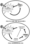
Figure 6. Model: Different Roles of RNAi Machinery in Somatic Mammalian Cells Versus Plants and Invertebrates
RNAi directly blocks viral replication in plants and invertebrate animals (top). RNAi and RISC components in somatic mammalian cells repress expression of ISG/antiviral effectors via miRNAs. RNAi is inhibited by activation of the intracellular antiviral response, allowing for optimal ISG expression, especially of cytotoxic ISGs (bottom).

References

    1. Backes S, Shapiro JS, Sabin LR, Pham AM, Reyes I, Moss B, Cherry S, tenOever BR. Degradation of host microRNAs by poxvirus poly(A) polymerase reveals terminal RNA methylation as a protective antiviral mechanism. Cell Host Microbe. 2012;12:200–210. - PMC - PubMed
    1. Barber GN. The interferons and cell death: guardians of the cell or accomplices of apoptosis? Semin Cancer Biol. 2000;10:103–111. - PubMed
    1. Bartel DP. MicroRNAs: target recognition and regulatory functions. Cell. 2009;136:215–233. - PMC - PubMed
    1. Bennasser Y, Le SY, Benkirane M, Jeang KT. Evidence that HIV-1 encodes an siRNA and a suppressor of RNA silencing. Immunity. 2005;22:607–619. - PubMed
    1. Chawla-Sarkar M, Lindner DJ, Liu YF, Williams BR, Sen GC, Silverman RH, Borden EC. Apoptosis and interferons: role of interferon-stimulated genes as mediators of apoptosis. Apoptosis. 2003;8:237–249. - PubMed

Publication types