. 2013 Oct 16;80(2):402-414.
doi: 10.1016/j.neuron.2013.07.046. Epub 2013 Sep 26.

Anti-tau antibodies that block tau aggregate seeding in vitro markedly decrease pathology and improve cognition in vivo

Affiliations

Anti-tau antibodies that block tau aggregate seeding in vitro markedly decrease pathology and improve cognition in vivo

Kiran Yanamandra et al. Neuron. .

Abstract

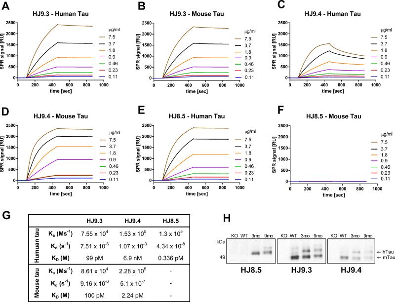
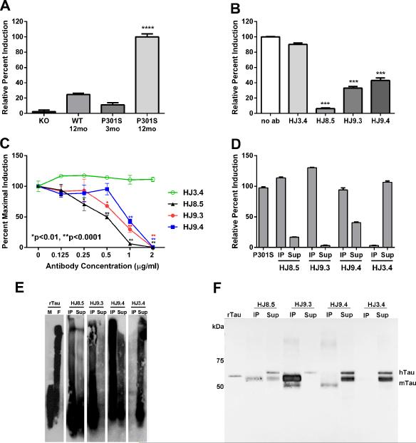
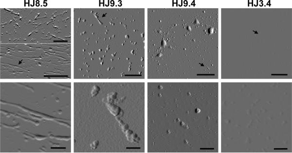
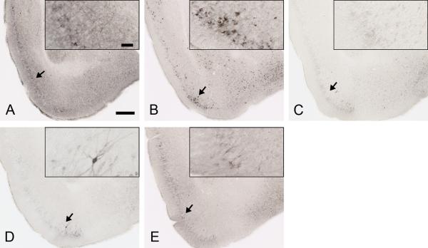
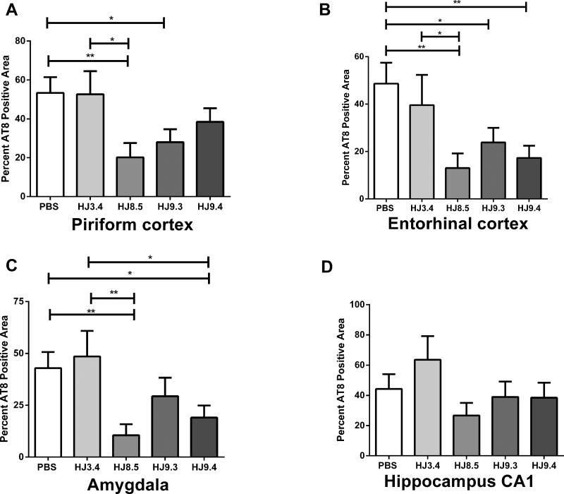
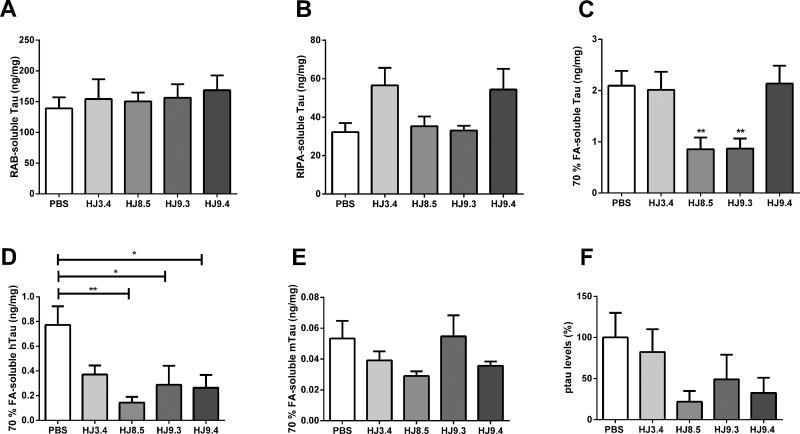
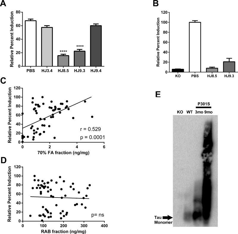
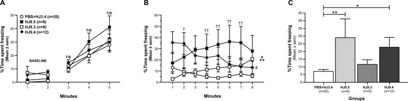
Tau aggregation occurs in neurodegenerative diseases including Alzheimer's disease and many other disorders collectively termed tauopathies. trans-cellular propagation of tau pathology, mediated by extracellular tau aggregates, may underlie pathogenesis of these conditions. P301S tau transgenic mice express mutant human tau protein and develop progressive tau pathology. Using a cell-based biosensor assay, we screened anti-tau monoclonal antibodies for their ability to block seeding activity present in P301S brain lysates. We infused three effective antibodies or controls into the lateral ventricle of P301S mice for 3 months. The antibodies markedly reduced hyperphosphorylated, aggregated, and insoluble tau. They also blocked development of tau seeding activity detected in brain lysates using the biosensor assay, reduced microglial activation, and improved cognitive deficits. These data imply a central role for extracellular tau aggregates in the development of pathology. They also suggest that immunotherapy specifically designed to block trans-cellular aggregate propagation will be a productive treatment strategy.


Figures

Figure 1
Figure 1. Characterization of anti-tau antibodies by surface plasmon resonance (SPR) and Immunoblotting
SPR sensorgrams showing the binding of each anti-tau antibody towards immobilized recombinant human tau (longest isoforms hTau40, 441 aa) and immobilized mouse tau (longest isoforms mTau40, 432 aa). Each antibody was run with various concentrations (0.11, 0.23, 0.46, 0.90, 1.8, 3.7, 7.5 μg/ml) and plots are shown in the corresponding color. (A) SPR sensorgrams of HJ9.3 antibody binding to immobilized human tau and immobilized mouse tau (B). (C) SPR sensorgrams of HJ9.4 antibody binding to immobilized human tau and immobilized mouse tau (D). SPR sensorgrams of HJ8.5 antibody binding to immobilized (E) human and (F) mouse tau. (G) Table showing the association rate constant (Ka), dissociation rate constant (Kd) and binding constant (KD) of each antibody towards human and mouse tau. BIAevaluation software (Biacore AB) was used to calculate Ka and Kd by selecting Fit kinetics simultaneous Ka/Kd (Global fitting) with 1:1 (Langmuir) interaction model. Ms−1=millisecond, M=molar, s=second. (H) RAB soluble fractions of 3 month old tau knockout (KO), 3 month old wild-type (WT), 3 month old P301S (3mo) and 9 month old P301S (9mo) mice were analyzed by immunoblot by using the indicated anti-tau antibodies.
Figure 2
Figure 2. Tau-antibodies block the uptake and seeding activity of P301S tau aggregates as detected by a FRET assay
HEK293 cells expressing RD (ΔK280)-CFP/YFP were exposed to 2.5 μg of total protein of 1xTBS brain lysates for 24 h. Brain lysates collected from 12 mo old P301S mice induced much greater seeding activity (n=5) as compared to lysates from knockout (KO) mice (n=7), wild type (WT) mice (n=6) or young 3-mo old P301S mice (n=2). ****p<0.0001 vs. other groups (B) HEK293 cells were co-transfected with RD (ΔK280)-CFP and RD (ΔK280)-YFP. 18hrs later, pre-incubated P301S brain lysates with or without incubation of anti-tau antibodies (HJ8.5, HJ9.3 and HJ9.4) or control antibody (HJ3.4, anti Aβ antibody) were added to cells. We found all the tau antibodies incubated with P301S brain lysates significantly blocked seeding activity. Statistical significance was determined by one-way ANOVA followed by Dunnett's post hoc test for multiple comparisons by using GraphPad Prism 5.0 software. ***p>0.001 (C) Titration of these antibodies with various concentrations (0.125 μg/ml, 0.25 μg/ml, 0.5 μg/ml, 1 μg/ml and 2 μg/ml) was performed with a fixed amount of P301S brain lysates. 24 hrs later, FRET analysis was performed. Out of all tau-antibodies we used, HJ8.5 was the most potent in blocking the uptake and seeding activity of P301S brain lysates. Statistical significance was determined by two-way ANOVA followed by Bonferroni post hoc test for multiple comparisons. ** p < 0.0001, * p < 0.01, Values represent mean ± SEM. Immunopecipitated (IP) and depleted supernatant (Sup) of P301S brain lysate of all anti tau-antibodies (HJ8.5, HJ9.3, HJ9.4) and control antibody (HJ3.4) was analyzed by tau seeding assay (D), SDD-AGE followed by western blot (E) and SDS-PAGE followed by western blot (F). Monomeric (M) and fibrillized (F) recombinant human tau (rTau) was used as controls for SDD-AGE and monomeric rTau was used for SDS-PAGE as a control. hTau indicates human tau and mTau indicates endogenous mouse tau.
Figure 3
Figure 3. AFM analysis of isolated tau aggregates from P301S mouse brain
Tau aggregates were isolated by immunoprecipitation (IP) from TBS lysates of 12 month old P301S mouse brains by using anti-tau antibodies (HJ8.5, HJ9.3, HJ9.4) and control antibody HJ3.4. Each column represents the IP material from each antibody. Black arrows indicate the areas magnified. Lower panel shows a magnified area of the upper panel. Scale bar in all upper panel images is 1 μm and in lower panel images is 200 nm. Morphology of the aggregated species IP'ed by each anti-tau antibody appears unique. Anti-Aβ antibody HJ3.4 did not IP any aggregates.
Figure 4
Figure 4. Anti-tau antibodies strongly decreased AT8 staining in P301S mouse brain
Representative coronal sections of PBS (A), HJ3.4 antibody (B), HJ8.5 antibody (C), HJ9.3 antibody (D) and HJ9.4 antibody (E) treated 9 month old P301S mice stained with biotinylated AT8 antibody in regions including the piriform cortex and amygdala. Scale bar is 250 μm. Inserts in A to E show the higher magnification of biotinylated AT8 antibody staining of phosphorylated tau, scale bar is 50 μm.
Figure 5
Figure 5. Certain anti-tau antibodies strongly decrease AT8 staining in P301S mouse brain
Percent of the area covered by biotinylated AT8 staining of abnormally phosphorylated tau in piriform cortex (A), entorhinal cortex (B), amygdala (C) and hippocampus CA1 region (D) in mice treated with the anti-tau antibodies HJ8.5 (N=13), HJ9.3 (N=15), HJ9.4 (N=13), the anti-Aβ antibody, HJ3.4 (N=8), or PBS (N=16) in 9 month old P301S mice. There was reduced AT8 staining in several different brain regions in the anti-tau antibody treated mice compared to PBS or HJ3.4 antibody treated mice. HJ8.5 had the largest effects. ** p < 0.01, * p < 0.05, values represent mean ± SEM.
Figure 6
Figure 6. Insoluble tau levels are reduced by antibodies HJ8.5 and HJ9.3 in P301S mice
The cortex of all the treated mice [PBS (N=16), HJ3.4 antibody (N=8) HJ8.5 (N=13), HJ9.3 (N=15), HJ9.4 (N=13)] were sequentially extracted by RAB (A), RIPA (B) and 70% FA (C) and their tau levels were quantified by ELISA. There were no statistical differences in soluble tau levels in RAB and RIPA fractions between the groups. However, there was a significant decrease of insoluble tau levels in 70% FA fractions in the HJ8.5 and HJ9.3 anti-tau antibodies treated mice compared to the PBS or HJ3.4 antibody treated groups. Insoluble tau levels in the HJ9.4 antibody treated mice were not different from the control groups. **p<0.01 Levels of human tau (D), mouse tau (E) and phospho tau at Ser202 and Thr205 (F) levels were assessed in 70% FA fractions by specific anti-human, anti-mouse, or anti-phospho tau antibodies by ELISA (n=6 mice per treatment group). There was a decrease in human tau levels in all groups of anti-tau antibody treated mice and no change in mouse tau levels. In 70% FA fractions, we also found that phospho tau at Ser202 and Thr205 as detected by AT8 reactivity was reduced in anti-tau antibody treated mice compared to controls, similar to total human tau.
Figure 7
Figure 7. Anti-tau antibody treated P301S mice have decreased tau seeding activity in cortical extracts as detected by FRET assay
(A) Tau seeding activity was measured with RAB soluble fractions of all PBS (N=16), HJ3.4 (N=8), HJ8.5 (N=13), HJ9.3 (N=15), and HJ9.4 (N=13) treated mice on HEK293 cells by FRET assay. HEK293 cells were co-transfected with RD (ΔK280)-CFP and RD (ΔK280)-YFP. 18 hrs later, RAB soluble fractions were added to cells. Seeding activity was significantly reduced in HJ8.5, and HJ9.3 antibody treated mice compared to the PBS or HJ3.4 antibody treated mice. RAB soluble fractions from HJ9.4 antibody treated mice did not have decreased seeding activity compared to the PBS or HJ3.4 antibody RAB soluble fractions. ***p<0.001, Values represent mean ± SEM. (B) RAB soluble fractions from tau KO, PBS-treated, and the anti-tau antibody treated mice were incubated with un-conjugated protein-G-agarose beads at 4°C with end-over-end rotation for 24 hours. This precipitates any residual antibody in the brain, including antibody bound to tau seeds. Elution of any seeding activity from the antibody/bead complexes was measured by FRET assay. There was significantly less seeding activity observed in HJ8.5 and HJ9.3 antibody treated mice versus PBS-treated mice ****p<0.0001, values represent mean ± SEM. (C) 70% FA fractions of 9 month old P301S brain cortex region of all treated groups analyzed by ELISA showed a strong correlation with FRET analysis performed with the RAB soluble fractions. (D) Comparison between tau levels (X-axis) and seeding activity (Y-axis) present in RAB soluble fractions of 9 month old P301S brain cortex of all treated mice assessed. There was no significant correlation between these 2 measures. (E) Tau species in the RAB soluble fractions of 3 month old knockout (KO), 3 month old wild type (WT), 3 month old P301S, and 9 month old PBS-treated P301S mice were separated on SDD-AGE, followed by western blotting. Polyclonal mouse anti-tau antibody was used for detecting tau species. High molecular weight tau species present in the RAB soluble fraction in both 3 month old P301S mice and larger amounts present in 9 month old P301S mice.
Figure 8
Figure 8. Contextual fear conditioning deficits in P301S tau transgenic mice are rescued by HJ8.5 and HJ9.4 antibody treatments
(A) On day 1 of conditioned fear testing, no differences were observed among groups in freezing levels during either the 2-min baseline condition or the tone/shock (T/S) training as indicated by the lack of a significant main or interaction effects involving Treatment following rmANOVAs on these data. (B) In contrast, a significant effect of Treatment (*p=0.019) and a significant Treatment by Minutes interaction (**p=0.0001) were observed following an rmANOVA on freezing levels during the contextual fear testing on day 2. Only the HJ9.4 group showed significant habituation from minute 1 versus minute 8, (#p=0.002). (C) Subsequent planned comparisons showed that freezing in the HJ8.5 and HJ9.4 tau antibody groups was significantly increased relative to the PBS+HJ3.4 control group when averaged across the 8-min session (**p=0.006 and *p=0.022, respectively). However, further analyses of the data showed that the largest differences between the HJ9.4 group and the PBS+HJ3.4 controls occurred during minute 2 (†p=0.004), while the largest differences between the HJ8.5 treated mice and the control group were found during minutes 4-7 (††p<0.004) as depicted in “B”.

Comment in

References

    1. Andoh T, Kuraishi Y. Direct action of immunoglobulin G on primary sensory neurons through Fc gamma receptor I. FASEB J. 2004;18:182–184. - PubMed
    1. Arriagada PV, Growdon JH, Hedley-Whyte ET, Hyman BT. Neurofibrillary tangles but not senile plaques parallel duration and severity of Alzheimer's disease. Neurology. 1992;42:631–639. - PubMed
    1. Asuni AA, Boutajangout A, Quartermain D, Sigurdsson EM. Immunotherapy targeting pathological tau conformers in a tangle mouse model reduces brain pathology with associated functional improvements. J Neurosci. 2007;27:9115–9129. - PMC - PubMed
    1. Bae EJ, Lee HJ, Rockenstein E, Ho DH, Park EB, Yang NY, Desplats P, Masliah E, Lee SJ. Antibody-aided clearance of extracellular alpha-synuclein prevents cell-to-cell aggregate transmission. J Neurosci. 2012;32:13454–13469. - PMC - PubMed
    1. Bancher C, Braak H, Fischer P, Jellinger KA. Neuropathological staging of Alzheimer lesions and intellectual status in Alzheimer's and Parkinson's disease patients. Neurosci Lett. 1993;162:179–182. - PubMed

Publication types

MeSH terms