. 2013 Oct 17;39(4):687-96.
doi: 10.1016/j.immuni.2013.08.019. Epub 2013 Sep 26.

Transforming growth factor-β signaling controls the formation and maintenance of gut-resident memory T cells by regulating migration and retention

Affiliations

Transforming growth factor-β signaling controls the formation and maintenance of gut-resident memory T cells by regulating migration and retention

Nu Zhang et al. Immunity. .

Abstract

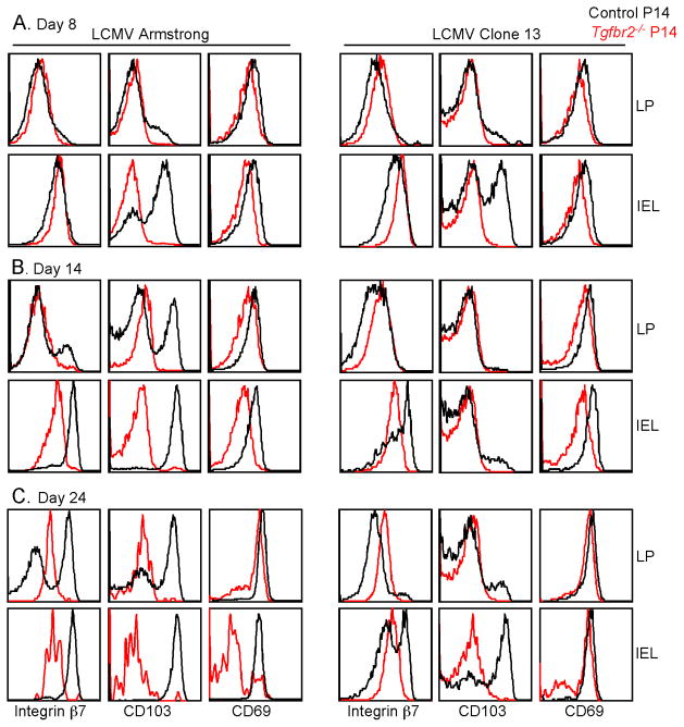
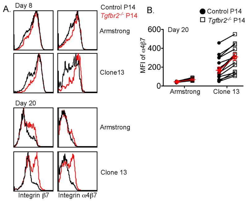
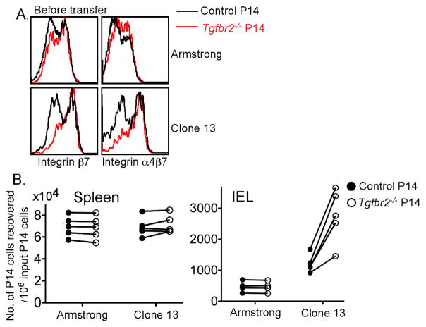
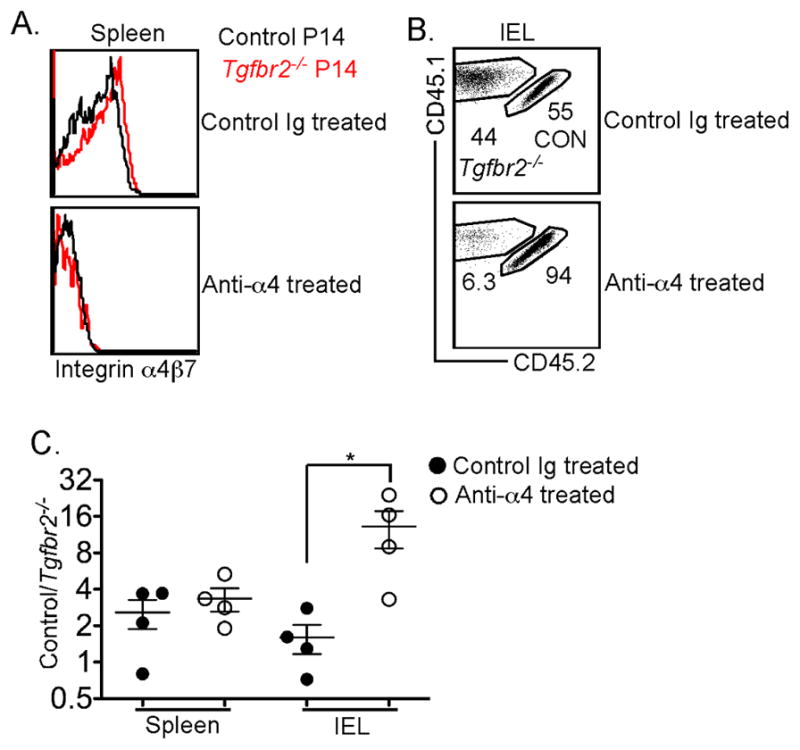
Tissue-resident memory T (Trm) cells represent a population of memory CD8⁺ T cells that can act as first responders to local infection. The mechanisms regulating the formation and maintenance of intestinal Trm cells remain elusive. Here we showed that transforming growth factor-β (TGF-β) controlled both stages of gut Trm cell differentiation through different mechanisms. During the formation phase of Trm cells, TGF-β signaling inhibited the migration of effector CD8⁺ T cells from the spleen to the gut by dampening the expression of integrin α4β7. During the maintenance phase, TGF-β was required for the retention of intestinal Trm cells at least in part through the induction of integrins αEβ7 and α1, as well as CD69. Thus, the cytokine acts to control cytotoxic T cell differentiation in lymphoid and peripheral organs.


Figures

Figure 1
Figure 1. Response in the spleen of TGF-βRII-deficient CD8+ T cells following acute and chronic LCMV infection
Naïve P14 T cells were purified from control (CD45.1+) and TGF-βRII-deficient (CD45.1+CD90.1+) mice, mixed at a 1:1 ratio, and 104 cells transferred into sex-matched B6 recipients followed by LCMV Armstrong or Clone 13 infection. The expression of TGF-βRII on P14 T cells is shown in A and B. Representative FACS plots of P14 T cells are shown in C. The ratio of control vs TGF-βRII-deficient P14 cells is shown in D (combined data from 4 independent experiments). Each symbol in B and D represents an individual recipient mouse. *, p<0.05 (Student t-test). Data in B and D are represented as mean +/− SEM. See also Figure S1.
Figure 2
Figure 2. Defective maintenance of TGF-βRII-deficient CD8+ T cells in the gut following acute, but not chronic LCMV infection
Similar experimental setup as in Figure 1. Data from LCMV Armstrong infected mice are shown in A, B and C, and from LCMV Clone 13 infected mice in D, E and F. At the indicated time post infection, lymphocytes were purified from the lamina propria and IEL compartment of the recipient mice. Representative FACS plots are shown in A and D. The ratio of control vs TGF-βRII-deficient P14 cells is shown in B and E (combined data from 4 independent experiments). The percentage of P14 T cells in CD8β+ IEL cells is shown in C and F (combined data from 4 independent experiments). Each symbol in B and E represents an individual recipient mouse. Data in B, C, E and F are represented as mean +/− SEM. See also Figure S2.
Figure 3
Figure 3. TGF-βRII-deficient cells in the gut exhibit reduced expression of markers associated with the retention
Similar experimental setup as in Figure 1. On day 8 (A), 14 (B) and 24 (C) post LCMV infection, lymphocytes were purified from the lamina propria and IEL compartment of the recipient mice. The expression of integrin β7, CD103 and CD69 on the P14 T cells were determined by flow cytometry. Data are representative of 4 independent experiments. See also Figure S3.
Figure 4
Figure 4. Defective retention of TGF-βRII-deficient T cells in the gut IEL
Similar experimental setup as in Figure 1. At day 8 and 12 post LCMV Arm infection and day 8 and 15 post LCMV Cl13 infection, IEL cells were purified following a two-step protocol. The ratio of control vs TGF-βRII-deficient P14 T cells in the tight and loose IEL populations is shown. Each pair of symbols represents an individual recipient mouse. Combined data from 3 independent experiments are shown. See also Figure S4.
Figure 5
Figure 5. Increased expression of integrin α4β7 on TGF-βRII-deficient CD8+ T cells in the spleen and peripheral blood
Similar experimental setup as in Figure 1. On day 8 and 20 post infection, the expression of integrin α4β7 and β7 by P14 T cells in the spleen (A) and peripheral blood (B) was examined. Each pair of symbols in (B) represents an individual recipient mouse. Red symbols represent the mean +/− SEM of 14–15 mice. Data are representative of 5 independent experiments. See also Figure S5.
Figure 6
Figure 6. Dramatically increased migration of TGF-βRII-deficient CD8+ T cells to the gut following LCMV Clone 13 infection
Similar experimental setup as in Figure 1. On day 9 post infection, 2x10⁷ pooled lymphocytes from spleen and lymph nodes were transferred into each naïve B6 mouse. 19 hours later, recipient mice were sacrificed and the number of transferred P14 T cells from different tissues was determined by flow cytometry and cell counting. (A) The expression of integrin α4β7 and β7 on the donor P14 T cells before secondary transfer. (B) The number of recovered P14 cells from the spleen and IEL compartment. Each pair of symbols represents an individual recipient mouse. Data are representative of two independent experiments. See also Figure S6.
Figure 7
Figure 7. Blocking integrin α4 results in defective maintenance of TGF-βRII-deficient CD8+ T cells in the gut following chronic LCMV infection
Naïve P14 T cells were purified from control (CD45.1+CD45.2+) and TGF-βRII-deficient (CD45.1+) mice, mixed at a 1:1 ratio, and 104 cells transferred into sex-matched B6 recipients followed by LCMV Clone 13 infection. Starting at day 10 post infection, 400μg anti-integrin α4 antibody (clone PS/2) or control Ig were injected intraperitoneally every other day (Figure S7A). The expression of integrin α4β7 on splenic P14 T cells is shown in A. Representative FACS profiles of the P14 T cells in the IEL compartment are shown in B. (C) The ratio of control vs Tgfbr2−/− P14 T cells in spleen and IEL is shown. Each symbol in C represents an individual mouse. *, p<0.05 (Student t-test). Data in C are represented as mean +/− SEM. See also Figure S7 and S8.

References

    1. Ahmed R, Salmi A, Butler LD, Chiller JM, Oldstone MB. Selection of genetic variants of lymphocytic choriomeningitis virus in spleens of persistently infected mice. Role in suppression of cytotoxic T lymphocyte response and viral persistence. J Exp Med. 1984;160:521–540. - PMC - PubMed
    1. Bargatze RF, Jutila MA, Butcher EC. Distinct roles of L-selectin and integrins alpha 4 beta 7 and LFA-1 in lymphocyte homing to Peyer’s patch-HEV in situ: the multistep model confirmed and refined. Immunity. 1995;3:99–108. - PubMed
    1. Berlin C, Bargatze RF, Campbell JJ, von Andrian UH, Szabo MC, Hasslen SR, Nelson RD, Berg EL, Erlandsen SL, Butcher EC. alpha 4 integrins mediate lymphocyte attachment and rolling under physiologic flow. Cell. 1995;80:413–422. - PubMed
    1. Berlin C, Berg EL, Briskin MJ, Andrew DP, Kilshaw PJ, Holzmann B, Weissman IL, Hamann A, Butcher EC. Alpha 4 beta 7 integrin mediates lymphocyte binding to the mucosal vascular addressin MAdCAM-1. Cell. 1993;74:185–195. - PubMed
    1. Boettler T, Cheng Y, Ehrhardt K, von Herrath M. TGF-beta blockade does not improve control of an established persistent viral infection. Viral Immunol. 2012;25:232–238. - PMC - PubMed

MeSH terms