Comparative Study
. 2013 Nov:116:227-33.
doi: 10.1016/j.exer.2013.09.007. Epub 2013 Sep 25.

Loss of the small heat shock protein αA-crystallin does not lead to detectable defects in early zebrafish lens development

Affiliations
Comparative Study

Loss of the small heat shock protein αA-crystallin does not lead to detectable defects in early zebrafish lens development

Mason Posner et al. Exp Eye Res. 2013 Nov.

Abstract

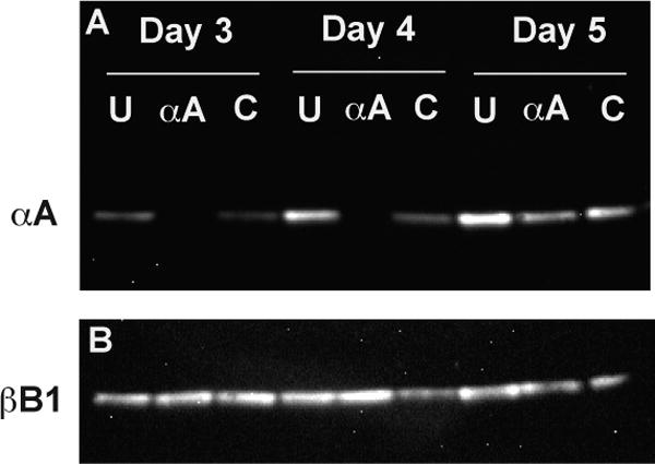
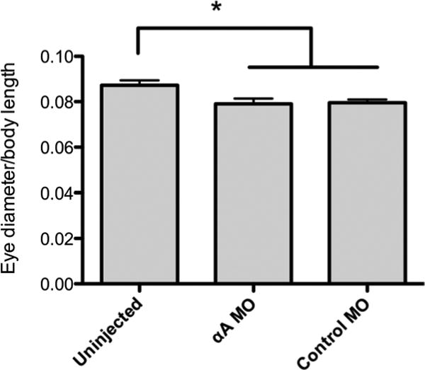
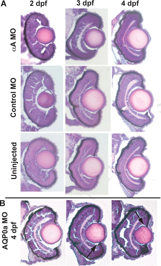
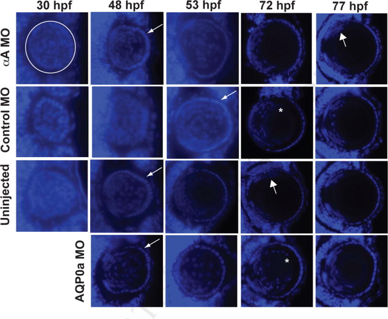
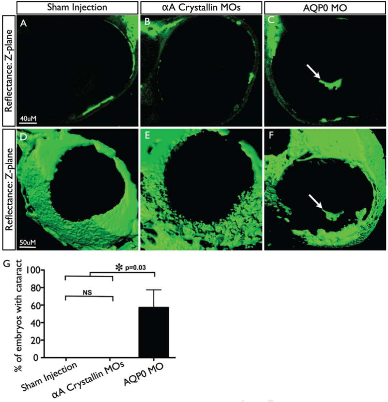
Alpha crystallins are small heat shock proteins essential to normal ocular lens function. They also help maintain homeostasis in many non-ocular vertebrate tissues and their expression levels change in multiple diseases of the nervous and cardiovascular system and during cancer. The specific roles that α-crystallins may play in eye development are unclear. Studies with knockout mice suggested that only one of the two mammalian α-crystallins is required for normal early lens development. However, studies in two fish species suggested that reduction of αA-crystallin alone could inhibit normal fiber cell differentiation, cause cataract and contribute to lens degeneration. In this study we used synthetic antisense morpholino oligomers to suppress the expression of zebrafish αA-crystallin to directly test the hypothesis that, unlike mammals, the zebrafish requires αA-crystallin for normal early lens development. Despite the reduction of zebrafish αA-crystallin protein to undetectable levels by western analysis through 4 days of development we found no changes in fiber cell differentiation, lens morphology or transparency. In contrast, suppression of AQP0a expression, previously shown to cause lens cataract, produced irregularly shaped lenses, delay in fiber cell differentiation and lens opacities detectable by confocal microscopy. The normal development observed in αA-crystallin deficient zebrafish embryos may reflect similarly non-essential roles for this protein in the early stages of both zebrafish and mammalian lens development. This finding has ramifications for a growing number of researchers taking advantage of the zebrafish's transparent external embryos to study vertebrate eye development. Our demonstration that lens cataracts can be visualized in three-dimensions by confocal microscopy in a living zebrafish provides a new tool for studying the causes, development and prevention of lens opacities.

Keywords: alpha crystallin; cataract; lens development; morpholino; ocular lens; small heat shock protein; zebrafish.


Figures

Fig. 1
Fig. 1
Alpha A-crystallin was undetectable by western analysis in MO knockdown embryos through four days post fertilization (dpf). Separate groups of 1-4 cell stage embryos were either left uninjected (U), or injected with morpholinos against αA-crystallin (αA) or a control morpholino (C) that does not recognize any zebrafish mRNA. Protein from the heads of embryos at three, four and five dpf were probed with antibodies against αA- and βB1-crystallin and detected with a chemiluminescent secondary antibody.
Fig. 2
Fig. 2
Knockdown of αA-crystallin did not affect eye size. Both αA-crystallin and control MO injected fish had smaller eyes as a ratio to body length compared to uninjected fish, but there was no significant difference between the αA-crystallin MO and control MO treated groups. Error bars indicate standard error of the mean (n=15). Asterisk indicates statistical significance at p < 0.05 (ANOVA with Tukey's multiple comparison test).
Fig. 3
Fig. 3
Knockdown of αA-crystallin produced no noticeable histological effects on lens development. A) Embryos on days two, three and four post fertilization were fixed, cryosectioned and stained with hematoxylin and eosin. These sections indicate no alterations in lens or retinal anatomy. Images are representative of at least three embryos examined for each timepoint, with none showing irregularities in lens development. B) Knockdown of AQP0a, which was already known to produce lens cataract by 3 dpf, produced a range of phenotypes including normal lenses and those with indentation (arrows) and persistent fiber cell nuclei. Three of six examined embryos showed some irregularities in lens development.
Fig. 4
Fig. 4
Knockdown of αA-crystallin did not inhibit fiber cell denucleation in comparison to control injected fish. Embryos were injected with either an MO that blocked the translation of αA-crystallin (αA MO), one that does not recognize any zebrafish mRNA sequence (Control MO) or one that blocks translation of a critical water channel (AQP0a MO). Other embryos were left uninjected. Embryos fixed at the specified times were cryosectioned and stained with DAPI to show cell nuclei. White circle in the upper left panel indicates the extent of the lens as a representative example with the cornea to the right. Thin arrows indicate first appearance of a noticeable lens epithelium as fiber cells become denucleated, thick arrows indicate first appearance of fiber cell nuclei restricted to a proliferative zone, and asterisks indicates residual fiber cell nuclei surrounding the lens nucleus. Times shown are hours post fertilization (hpf). At least three embryos were examined for all timepoints, except for the 77 hour timepoint, for which 2 embryos were examined.
Figure 5
Figure 5
αA crystallin knockdown does not induce cataract formation. Representative z-plane (A-C) and 3D projections (D-F) of reflectance confocal image series taken of embryos injected with buffer only (A,D), αA crystallin MOs (B,E) or AQP0a MO (Positive control) (C,F). Arrows indicate cataracts, seen only in AQP0a injected MOs. (G) Percentage of embryos with cataracts (only one eye imaged per embryo), n≥7 for each condition. Error bars indicate standard deviation; two-tailed t-test used to measure statistical significance.

References

    1. Andley UP. Effects of alpha-crystallin on lens cell function and cataract pathology. Curr Mol Med. 2009;9:887–892. - PubMed
    1. Bhat SP. Transparency and non-refractive functions of crystallins--a proposal. Exp Eye Res. 2004;79:809–816. - PubMed
    1. Bhat SP, Nagineni CN. alpha B subunit of lens-specific protein alpha-crystallin is present in other ocular and non-ocular tissues. Biochem Biophys Res Commun. 1989;158:319–325. - PubMed
    1. Boyle DL, Takemoto LJ. A possible role for alpha-crystallins in lens epithelial cell differentiation. Mol Vis. 2000;6:63–71. - PubMed
    1. Boyle DL, Takemoto LJ, Brady JP, Wawrousek EF. Morphological characterization of the Alpha A- and Alpha B-crystallin double knockout mouse lens. BMC Ophthalmology. 2003;3:3. - PMC - PubMed

Publication types

MeSH terms

LinkOut - more resources