

Hypoxia-induced expression of VEGF splice variants and protein in four retinal cell types
- PMID: 24076411
- PMCID: PMC4256053
- DOI: 10.1016/j.exer.2013.09.014
Hypoxia-induced expression of VEGF splice variants and protein in four retinal cell types
Abstract
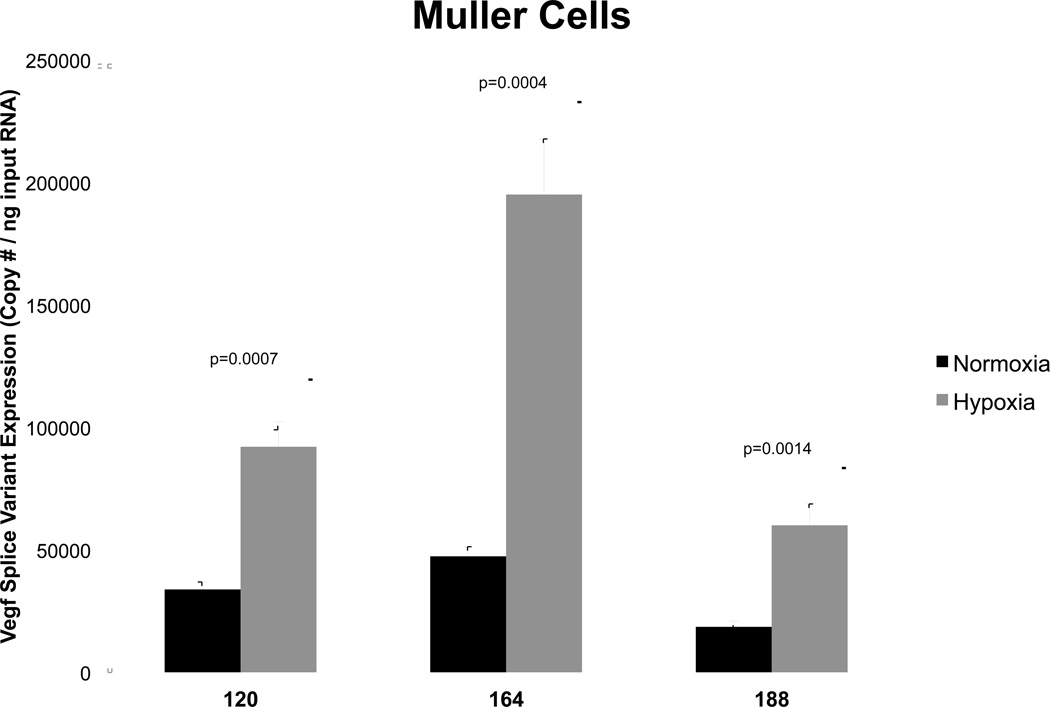
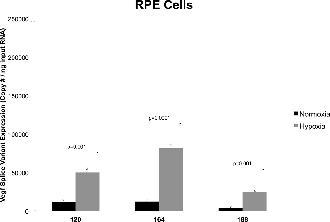
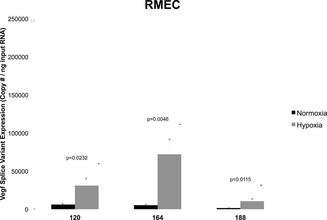
The purpose of this study was to investigate the hypoxia-induced Vegf120, Vegf164 and Vegf188 mRNA expression profiles in rat Müller cells (MC), astrocytes, retinal pigmented epithelial cells (RPE) and retinal microvascular endothelial cells (RMEC) and correlate these findings to VEGF secreted protein. Cultured cells were exposed to normoxia or hypoxia. Total RNA was isolated from cell lysates and Vegf splice variant mRNA copy numbers were assayed by a validated qRT-PCR external calibration curve method. mRNA copy numbers were normalized to input total RNA. Conditioned medium was collected from cells and assayed for total VEGF protein by ELISA. Hypoxia increased total Vegf mRNA and secreted protein in all the retinal cell types, with the highest levels observed in MC and astrocytes ranking second. Total Vegf mRNA levels in hypoxic RPE and RMEC were comparable; however, the greatest hypoxic induction of each Vegf splice variant mRNA was observed in RMEC. RPE and RMEC ranked 3rd and 4th respectively, in terms of secreted total VEGF protein in hypoxia. The Vegf120, Vegf164 and Vegf188 mRNA splice variants were all increased in hypoxic cells compared to normoxic controls. In normoxia, the relative Vegf splice variant mRNA levels ranked from highest to lowest for each cell type were Vegf164 > Vegf120 > Vegf188. Hypoxic induction did not alter this ranking, although it did favor an increased stoichiometry of Vegf164 mRNA over the other two splice variants. MC and astrocytes are likely to be the major sources of total Vegf, Vegf164 splice variant mRNAs, and VEGF protein in retinal hypoxia.
Keywords: hypoxia; splice variants; vascular endothelial growth factor.
Copyright © 2013 Elsevier Ltd. All rights reserved.
Figures


References
-
- Aiello LP, Avery RL, et al. Vascular endothelial growth factor in ocular fluid of patients with diabetic retinopathy and other retinal disorders. The New England journal of medicine. 1994;331(22):1480–1487. - PubMed
-
- Aiello LP, Northrup JM, et al. Hypoxic regulation of vascular endothelial growth factor in retinal cells. Archives of ophthalmology. 1995;113(12):1538–1544. - PubMed
-
- Bai Y, Ma JX, et al. Muller cell-derived VEGF is a significant contributor to retinal neovascularization. The Journal of pathology. 2009;219(4):446–454. - PubMed
-
- Ben-Av P, Crofford LJ, et al. Induction of vascular endothelial growth factor expression in synovial fibroblasts by prostaglandin E and interleukin-1: a potential mechanism for inflammatory angiogenesis. FEBS letters. 1995;372(1):83–87. - PubMed
Publication types
MeSH terms
Substances
Grants and funding
LinkOut - more resources
Full Text Sources
Other Literature Sources
Medical
