. 2013 Nov;45(11):1386-91.
doi: 10.1038/ng.2777. Epub 2013 Sep 29.

Global chromatin profiling reveals NSD2 mutations in pediatric acute lymphoblastic leukemia

Affiliations

Global chromatin profiling reveals NSD2 mutations in pediatric acute lymphoblastic leukemia

Jacob D Jaffe et al. Nat Genet. 2013 Nov.

Abstract

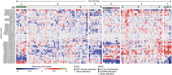
Epigenetic dysregulation is an emerging hallmark of cancers. We developed a high-information-content mass spectrometry approach to profile global histone modifications in human cancers. When applied to 115 lines from the Cancer Cell Line Encyclopedia, this approach identified distinct molecular chromatin signatures. One signature was characterized by increased histone 3 lysine 36 (H3K36) dimethylation, exhibited by several lines harboring translocations in NSD2, which encodes a methyltransferase. A previously unknown NSD2 p.Glu1099Lys (p.E1099K) variant was identified in nontranslocated acute lymphoblastic leukemia (ALL) cell lines sharing this signature. Ectopic expression of the variant induced a chromatin signature characteristic of NSD2 hyperactivation and promoted transformation. NSD2 knockdown selectively inhibited the proliferation of NSD2-mutant lines and impaired the in vivo growth of an NSD2-mutant ALL xenograft. Sequencing analysis of >1,000 pediatric cancer genomes identified the NSD2 p.E1099K alteration in 14% of t(12;21) ETV6-RUNX1-containing ALLs. These findings identify NSD2 as a potential therapeutic target for pediatric ALL and provide a general framework for the functional annotation of cancer epigenomes.


Figures

Figure 1
Figure 1
Global chromatin profiling identifies distinct molecular chromatin-signature profiles in the CCLE collection. We subjected 115 cell lines to molecular chromatin-signature profiling by MS. Each row corresponds to a H3 peptide with the specific combination of marks shown (left). The value in each cell of the heatmap corresponds to the log2-fold change of the mark combination versus the median for each row. EZH2 and NSD2 status are indicated. Clusters A–F are identified for reference in the main text.
Figure 2
Figure 2
H3K27 and H3K36 methylation alterations drive the identification of unique molecular chromatin signatures. Molecular chromatin-signature profiles of clusters A, D and F, magnified to show a subset of histone H3 peptides that drive the key signatures of these three clusters.
Figure 3
Figure 3
Identification of recurrent NSD2 alterations in ALL. (a) Distribution of NSD2 p.E1099K cell lines across 181 CCLE cell lines of hematopoietic origin. AML, acute myeloid leukemia; CML, chronic myeloid leukemia; MCL, mantle-cell lymphoma; BL, Burkitt’s lymphoma; DLBCL, diffuse large B-cell lymphoma; HL, Hodgkin’s lymphoma. Six ALL cell lines and one myeloma cell line contain the p.E1099K alteration (red). Eight myeloma cell lines contain the t(4;14) translocation (gray). (b) Mapping of the NSD2 p.E1099K alteration on the domain structure of NSD2. Bottom, alignment of amino acids surrounding E1099 in the SET domain sequence of NSD2 with other histone methyltransferases. NSD1, NSD2, NSD3, SETD2 and MES4 methylate H3K36. Boxes indicate identical residues among NSD proteins; red dots indicate identical residues across all sequences. (c) Structural modeling of NSD2 E1099 (magenta stick) to K1099 (white stick) alteration. It is predicted to alter binding between NSD2 (shown in green) and the histone peptide (shown in blue). C1183 (green stick), S31 (blue stick) and K36 (blue stick) are shown to identify modeled locations between residues on NSD2 and the H3 peptide. Dashed line indicates interactions between NSD2 and H3 peptide.
Figure 4
Figure 4
NSD2 p.E1099K alteration leads to increased enzymatic activity and promotes transformation. (a) Biochemical characterization of the enzymatic activity of wild-type (WT) and p.E1099K-mutant NSD2. The catalytic domain of NSD2 (955–1365) was purified and assayed with nucleosome substrates. Enzymatic activity was assessed by quantifying the production of S-adenosyl-L-homocysteine (SAH) on the y axis. Error bars indicate the s.d. of triplicate measurements. (b) Western blot analysis of lysates from KMS11-TKO cells reconstituted with WT NSD2, p.E1099K-mutant NSD2, CDM NSD2 or both p.E1099K and CDM (p.E1099K-CDM) NSD2 or empty vector, analyzed with the indicated antibodies. Histone H3, loading control. (c) Quantification of soft-agar transformation (5% serum) of KMS11-TKO cells reconstituted with the indicated NSD2 variants. Error bars indicate the s.d. of triplicate samples (see also Supplementary Fig. 5). (d) Global chromatin profiling of KMS11-TKO engineered lines. Chromatin-signature profiling, clustering and color coding are as described in Figure 1. KMS11-TKO cells were reconstituted with the indicated NSD2 variants. The chromatin-signature profiling of KMS11-TKO reconstituted cells is shown with a representative panel of hematopoietic cell lines (see Supplementary Fig. 4 for complete data set and dendrogram shown).
Figure 5
Figure 5
NSD2 is required for the growth and proliferation of ALL cells carrying the p.E1099K alteration. (a) Immunoblots of NSD2, H3K36me2, H3 and γ-tubulin in a panel of six leukemic cell lines (red text indicates NSD2 1099K mutant cell lines). JVM-2 is B-cell chronic lymphocytic leukemia cell line; and the remaining five samples are from ALL cell lines. Doxycycline (dox) was used to induce the expression of two independent NSD2 shRNAs (sh1 and sh2). (b) Cell-proliferation assays of the indicated cell lines after NSD2 knockdown. The bars represent proliferation in dox-treated cells relative to untreated controls after 12 d. Error bars indicate s.d. of duplicate samples. Red text indicates NSD2 1099K mutant cell lines. (c) Soft-agar colony formation assay of SEM cells expressing the indicated shRNAs. (d) Quantification of soft-agar colony formation of the indicated ALL cell lines in response to induction (+ dox) of the indicated shRNAs. GFP shRNA was included as control (con). Error bars indicate the s.d. of triplicate samples. (e) Effect of inducible NSD2 and GFP shRNAs in the presence and absence of dox on the growth of subcutaneous SEM xenografts. Mean tumor volume ± s.e.m. is shown (n = 6 for NSD2 shRNA; n = 5 for GFP shRNA). (f) Immunoblots of NSD2 and H3K36me2 of tumor xenografts 18 d after dox induction. Individual tumor samples from each treatment group are shown (n = 6 for NSD2 shRNA; n = 5 for GFP shRNA). H3 and γ-tubulin were included as loading controls.

Comment in

References

    1. Barretina J, et al. The Cancer Cell Line Encyclopedia enables predictive modelling of anticancer drug sensitivity. Nature. 2012;483:603–607. - PMC - PubMed
    1. Peach SE, Rudomin EL, Udeshi ND, Carr SA, Jaffe JD. Quantitative assessment of chromatin immunoprecipitation grade antibodies directed against histone modifications reveals patterns of co-occurring marks on histone protein molecules. Mol. Cell. Proteomics. 2012;11:128–137. - PMC - PubMed
    1. Ryan RJ, Bernstein BE. Molecular biology. Genetic events that shape the cancer epigenome. Science. 2012;336:1513–1514. - PubMed
    1. Morin RD, et al. Frequent mutation of histone-modifying genes in non-Hodgkin lymphoma. Nature. 2011;476:298–303. - PMC - PubMed
    1. Shih AH, Abdel-Wahab O, Patel JP, Levine RL. The role of mutations in epigenetic regulators in myeloid malignancies. Nat. Rev. Cancer. 2012;12:599–612. - PubMed

Publication types