. 2013 Oct;8(10):735-41.
doi: 10.1038/nnano.2013.194. Epub 2013 Sep 29.

Sensitive capture of circulating tumour cells by functionalized graphene oxide nanosheets

Affiliations

Sensitive capture of circulating tumour cells by functionalized graphene oxide nanosheets

Hyeun Joong Yoon et al. Nat Nanotechnol. 2013 Oct.

Abstract

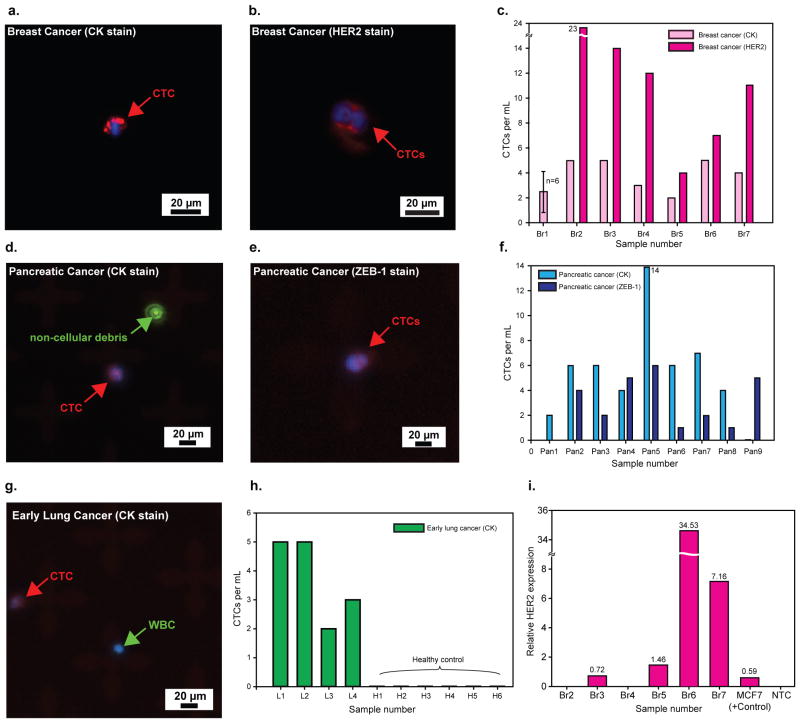
The spread of cancer throughout the body is driven by circulating tumour cells (CTCs). These cells detach from the primary tumour and move from the bloodstream to a new site of subsequent tumour growth. They also carry information about the primary tumour and have the potential to be valuable biomarkers for disease diagnosis and progression, and for the molecular characterization of certain biological properties of the tumour. However, the limited sensitivity and specificity of current methods for measuring and studying these cells in patient blood samples prevents the realization of their full clinical potential. The use of microfluidic devices is a promising method for isolating CTCs. However, the devices are reliant on three-dimensional structures, which limits further characterization and expansion of cells on the chip. Here we demonstrate an effective approach to isolating CTCs from blood samples of pancreatic, breast and lung cancer patients, by using functionalized graphene oxide nanosheets on a patterned gold surface. CTCs were captured with high sensitivity at a low concentration of target cells (73 ± 32.4% at 3-5 cells per ml blood).


Figures

Figure 1
Figure 1. GO chip and functionalisation/characterisation of GO
a, Schematic diagram of the GO chip. b, Schematic showing the conjugation chemistry between functionalised GO nanosheets and EpCAM antibodies. GO nanosheets are adsorbed onto the gold pattern. The GMBS crosslinker binds to PL-PEG-NH2 onto the GO nanosheets. The NeutrAvidin is connected to the GMBS and biotinylated EpCAM. c, Preparation procedures of the functionalized GO. d, SEM image of gold patterns. Inset: magnified SEM image of adsorbed GO nanosheets on gold patterns.
Figure 2
Figure 2. Characterization of the GO-chip with cells in buffer solution
a, Flow rate dependency on capture efficiency of MCF-7 cells. Error bars represent the standard deviation of three replicates. b, Capture efficiency of MCF-7 cells at the 1 mL/hr. The red solid line is a fit to the result expected. c, Comparison of different cell lines; MCF-7 cells (high EpCAM expression), PC-3 cells (low EpCAM expression), and Hs-578T cells (no EpCAM expression). Error bars represent the standard deviation of three replicates. d, Cell recovery of MCF-7 cells compared to functionalized flat silicon device. Error bars represent the standard deviation of three replicates. Inset: photograph of GO chip (left) and photograph of functionalized flat silicon device (right). e, Fluorescence image of the captured MCF-7 cells. Inset: magnified (10x) fluorescence image of the captured MCF-7 cells. f, SEM image of the captured MCF-7 cell on the gold pattern. Inset: magnified SEM image of the captured MCF-7 cell.
Figure 3
Figure 3. Characterization of the GO-chip with MCF-7 cells spiked into whole blood
a, Cell recovery MCF-7 cells spiked into 1 mL of whole blood at varying spike concentration from 3 to 100 cells/mL. Error bars represent the standard deviation of replicates. b, Fluorescence microscope image of MCF-7 and white blood cells stained with DAPI (blue), cytokeratin (red), and CD 45 (green). c, Fluorescence microscope image of 6-day-cultured MCF-7 cells. d, Fluorescence microscope image depicting proliferation of cells. e, SEM image of a captured and 6-day-cultured MCF-7 cell.
Figure 4
Figure 4. Fluorescence microscope images and quantification of CTCs captured from cancer patient samples
a, CTC captured from breast cancer patient #6 (Br6). b, CTCs captured from breast cancer patient #2 (Br2). c, Quantification of CTCs captured from breast cancer patients. d, CTC captured from pancreatic cancer patient #2 (Pan2); Fluorescence particle from Alexa Fluor 488 dye shows that CTC has no FITC (green). e, Two CTCs captured from pancreatic cancer patient #9 (Pan9). f, Quantification of CTCs captured from pancreatic cancer patients. g, CTC captured from early lung cancer patient #3 (L3). h, Quantification of CTCs captured from lung cancer patients and 6 healthy donors. i, HER2 gene expression (normalized to GAPDH) of captured CTCs by qRT-PCR. Four out of six patients showed expression higher than the gene expression of MCF-7 cells (100 cells).

Comment in

References

    1. Cristofanilli M, et al. Circulating Tumor Cells, Disease Progression, and Survival in Metastatic Breast Cancer. New England Journal of Medicine. 2004;351:781–791. - PubMed
    1. Nagrath S, et al. Isolation of rare circulating tumour cells in cancer patients by microchip technology. Nature. 2007;450:1235–1239. - PMC - PubMed
    1. Maheswaran S, et al. Detection of Mutations in EGFR in Circulating Lung-Cancer Cells. New England Journal of Medicine. 2008;359:366–377. - PMC - PubMed
    1. Willipinski-Stapelfeldt B, et al. Changes in Cytoskeletal Protein Composition Indicative of an Epithelial-Mesenchymal Transition in Human Micrometastatic and Primary Breast Carcinoma Cells. Clinical Cancer Research. 2005;11:8006–8014. - PubMed
    1. Bednarz-Knoll N, Alix-Panabières C, Pantel K. Plasticity of disseminating cancer cells in patients with epithelial malignancies. Cancer and Metastasis Reviews. 2012;31:673–687. - PubMed

Publication types