. 2013 Oct;8(10):763-71.
doi: 10.1038/nnano.2013.190. Epub 2013 Sep 29.

Self-assembly of carbon nanotubes and antibodies on tumours for targeted amplified delivery

Affiliations

Self-assembly of carbon nanotubes and antibodies on tumours for targeted amplified delivery

J Justin Mulvey et al. Nat Nanotechnol. 2013 Oct.

Abstract

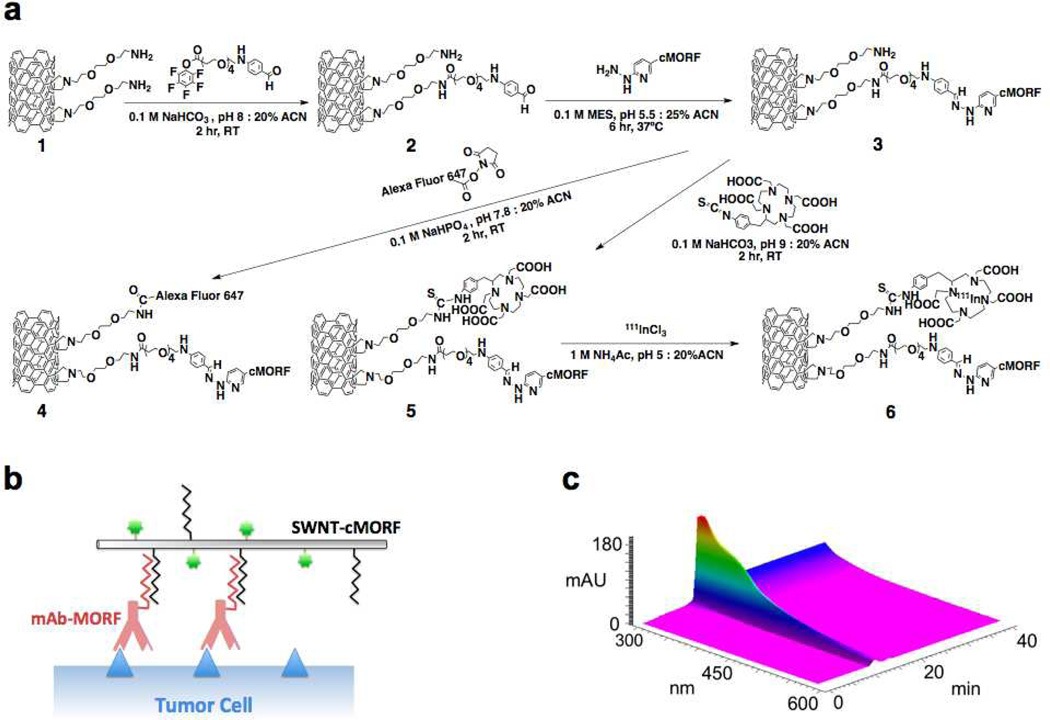
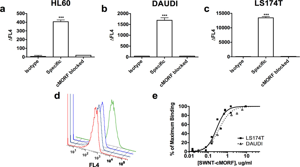
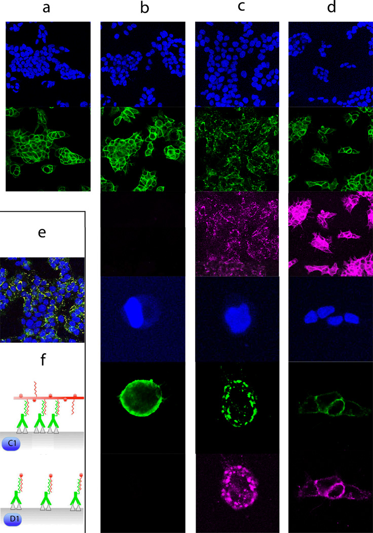
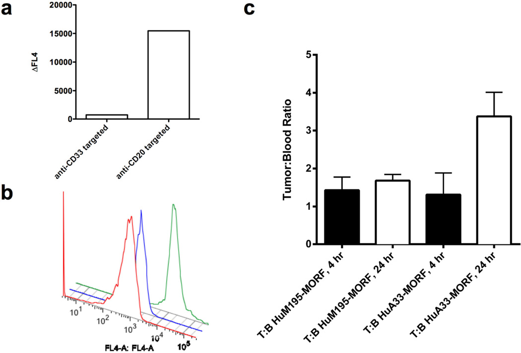
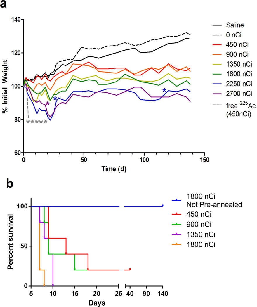
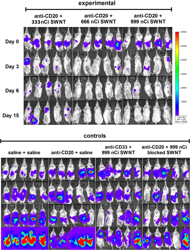
Single-walled carbon nanotubes (SWNTs) can deliver imaging agents or drugs to tumours and offer significant advantages over approaches based on antibodies or other nanomaterials. In particular, the nanotubes can carry a substantial amount of cargo (100 times more than a monoclonal antibody), but can still be rapidly eliminated from the circulation by renal filtration, like a small molecule, due to their high aspect ratio. Here we show that SWNTs can target tumours in a two-step approach in which nanotubes modified with morpholino oligonucleotide sequences bind to cancer cells that have been pretargeted with antibodies modified with oligonucleotide strands complementary to those on the nanotubes. The nanotubes can carry fluorophores or radioisotopes, and are shown to selectively bind to cancer cells in vitro and in tumour-bearing xenografted mice. The binding process is also found to lead to antigen capping and internalization of the antibody-nanotube complexes. The nanotube conjugates were labelled with both alpha-particle and gamma-ray emitting isotopes, at high specific activities. Conjugates labelled with alpha-particle-generating (225)Ac were found to clear rapidly, thus mitigating radioisotope toxicity, and were shown to be therapeutically effective in vivo.


Figures

Figure 1
Figure 1. Design and HPLC characterization of self-assembling SWNT-cMORF constructs
(a) Synthetic scheme for chemical functionalization of SWNT-NH2 to produce radio- and fluorescent labeled SWNT-cMORF conjugates (b) Diagrammatic representation (not to scale) of SWNT-cMORF self-assembly onto mAb-MORF targeted tumour cells. Blue triangle is tumour antigen; green dot is appended cytotoxic or diagnostic moiety. (c) 3D gel permeation HPLC chromatograph of compound 3 (SWNT-cMORF-NH2) tracing the spectrum of eluted material across time. Only a single peak is evident and the spectrum is consistent with SWNT modified with bis-aryl hydrazone groups (shoulder at λ = ~354 nm and decreasing absorption through 600nm – an identifying quality of nanotubes)
Figure 2
Figure 2. Self-assembly of SWNT-cMORF onto tumour cells pretargeted with mAb-MORF is highly specific and high affinity
a) Quantitation of flow cytometric assay of binding of SWNT-cMORF-AF647 onto HL60 cells. Pretreatment was with HL60 specific anti-CD33-MORF, isotype control anti-CD20-MORF, or specific anti-CD33-MORF + blocking with excess free cMORF. Data is presented as the change in median fluorescence. (b) Quantitation of flow cytometric assay of binding of SWNT-cMORF-AF647 onto DAUDI cells. Pretreatment was with DAUDI specific anti-CD20-MORF, isotype control anti-CD33-MORF, or specific anti-CD20-MORF + blocking with excess free cMORF. (c) Quantitation of flow cytometric assay of binding of SWNT-cMORF-AF647 onto LS174T cells. Pretreatment was with LS174T specific anti-A33-MORF, isotype control anti-CD33-MORF, or specific anti-A33-MORF + blocking with excess free cMORF. (d) Representative flow cytometric histogram of cell binding assay with SWNT-cMORF-AF647 on untreated (red), isotype control pretargeted (black), specific pretargeted + cMORF block (blue), or specific pretargeted (green) LS174T cells. (e) Binding curve for SWNT-cMORF-AF647 onto anti-CD20 pretargeted Daudi cells (squares) and anti-A33 pretargeted LS174T cells (triangles). Curves were fit using GraphPad Prism using an algorithm for one-site specific binding with variable Hill slope (R2 = 0.95, 0.97).
Figure 3
Figure 3. SWNT-cMORF conjugates are able to induce antigen capping and internalization when self-assembled onto cells targeted with mAb-MORF (images organized in columns)
(a) Anti-A33 (green, row 2) remained stably bound to LS174T cells (blue, DAPI nuclear stain, row 1) at 37°C for up to 24 hours (4 hours is shown) and were evenly distributed about the cell membrane. (b) Anti-A33-MORF conjugates (green, row 2,5) exhibited similar surface stability when bound to LS174T cells (blue, rows 1,3). High power images are provided (rows 4,5,6), as well 647nm views showing the absence signal (rows 3,6) (c) Cells pretreated with A33-MORF (green, rows 2,5) followed by SWNT-cMORF-AF647 (pink, rows 3,6), allowing for self-assembly at 37°C for up to 4 hours, demonstrated a significant change in the distribution of the bound antibody with a change to scattered punctate staining. A similar pattern was observed for 30 min and 1 hr incubations. High power images are provided (rows 4,5,6). (d) Cells treated with anti-A33-MORF (green, rows 2,5) followed by cMORF-AF647 (pink, rows 3,6) alone did not demonstrate change in distribution of A33 about the cell membrane. High power images are provided (rows 4,5,6). Anti-A33-MORF was evenly distributed about the plasma membrane, and cMORF-AF647 co-localized with the targeted mAb-MORF. (e) A composite staining of conditions in (c) with pink and green forming a yellow overlay (enlargement provided in Figure S2). (f) Schematic model of self-assembly process and cross-linking shown (not to scale) illustrating conditions of columns (c), above and (d).
Figure 4
Figure 4. SWNT-cMORF can selectively self-assemble onto pretargeted tumours in-vivo
(a) Flow cytometric analysis of CD20 positive, CD33 negative Daudi-GFP cells harvested from mice treated with either isotype control anti-CD33-MORF, or tumour specific anti-CD20-MORF. Data show the shift in median fluorescence from untreated Daudi-GFP cells (representative mice are shown). The SWNT-cMORF bound selectively to the tumour cells in mice pre-targeted with specific antibody as evidenced by the 20-fold increase over the control. (b) Representative flow cytometric histograms of data in panel (a) shows untreated cells (red), isotype mAb-MORF (anti-CD33-MORF) treated cells (blue), and specific mAb-MORF (anti-CD20-MORF) treated cells (green). (c) Tumour to blood ratios (T:B) for LS174T tumoured mice pretargeted with either specific anti-A33-MORF or isotype control anti-CD33-MORF followed by injection of SWNT-cMORF-111In(DOTA). Signal measurements in %ID per gram were measured at 4 or 24 hours post injection of injection of SWNT-cMORF-111In(DOTA). There was a significant increase (p<0.05) in tumour accumulation of SWNT-cMORF-111In(DOTA) at the tumour site only when pre-targeted with specific antibody as compared to isotype control.
Figure 5
Figure 5. SWNT-cMORF-(225Ac)DOTA mitigates radioisotope toxicity and can be used as an effective agent in multistep therapy of disseminated lymphoma
(a) Whole animal weights of tumour-free mice treated with varying dose levels of 225Ac labeled SWNT-cMORF-DOTA. An additional group of mice received 450 nCi of free 225Ac as a control. Data reflect average weights, n=5 for all groups. Each animal death is noted by a single asterisk. Toxicity study was halted at 140 days. Median survival was greater than 20 weeks in all groups (b) Kaplan-Meier plot of pre-annealed “single-step” treatment mouse survival with comparison to not annealed (a) 1800 nCi group from Fig 5a.. Toxicity study was halted at 40 days. Median survival was less than 2 weeks in all groups.
Figure 6
Figure 6. SWNT-cMORF-(225Ac)DOTA can be used as an effective agent in multistep therapy of disseminated lymphoma
Mice previously implanted with luciferase-transfected Daudi lymphoma cells were injected with 5mg/ml luciferin and imaged after a 5 minute delay. The parameters are equivalent for all images, but as a trade-off, this leads to saturation that disallows quantification. Mice were treated with multistep therapy as previously noted and imaged at days 0, 3, 6, and 15 post treatment. Growth, Rituximab therapy, isotype high-dose radiation, and blocked 2-step controls are provided. For further controls see also Figure S8.

Comment in

References

    1. Scheinberg DA, Villa CH, Escorcia FE, McDevitt MR. Conscripts of the infinite armada: systemic cancer therapy using nanomaterials. Nat. Rev. Clin. Oncol. 2010;7:266–276. - PMC - PubMed
    1. Davis ME, Chen ZG, Shin DM. Nanoparticle therapeutics: an emerging treatment modality for cancer. Nat. Rev. Drug Discov. 2008;7:771–782. - PubMed
    1. Kostarelos K, Bianco A, Prato M. Promises, facts and challenges for carbon nanotubes in imaging and therapeutics. Nature Nanotech. 2009;4:627–633. - PubMed
    1. Liu Z, et al. Drug delivery with carbon nanotubes for in vivo cancer treatment. Cancer research. 2008;68:6652–6660. - PMC - PubMed
    1. Liu Z, et al. In vivo biodistribution and highly efficient tumour targeting of carbon nanotubes in mice. Nature Nanotech. 2007;2:47–52. - PubMed

Publication types