. 2013 Sep 26;14(10):19540-55.
doi: 10.3390/ijms141019540.

Isolation and characterization of squamous cell carcinoma-derived stem-like cells: role in tumor formation

Affiliations

Isolation and characterization of squamous cell carcinoma-derived stem-like cells: role in tumor formation

Katiuscia Dallaglio et al. Int J Mol Sci. .

Abstract

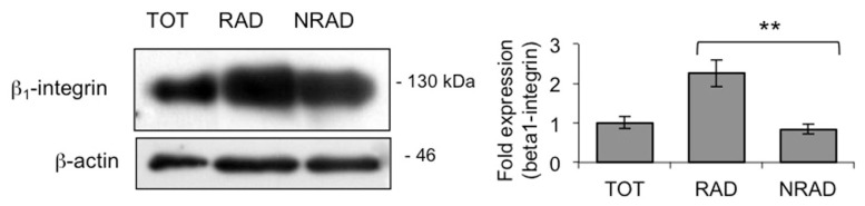
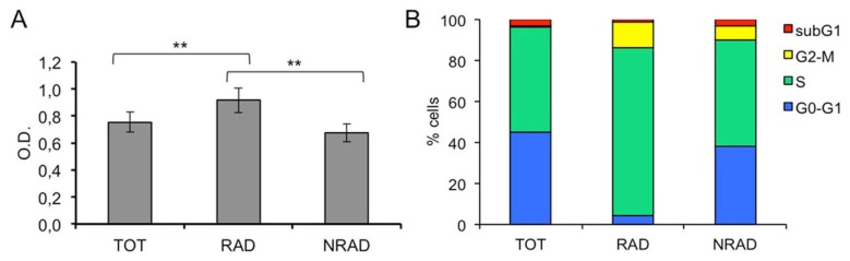
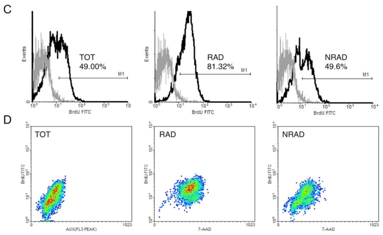
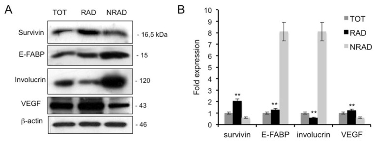
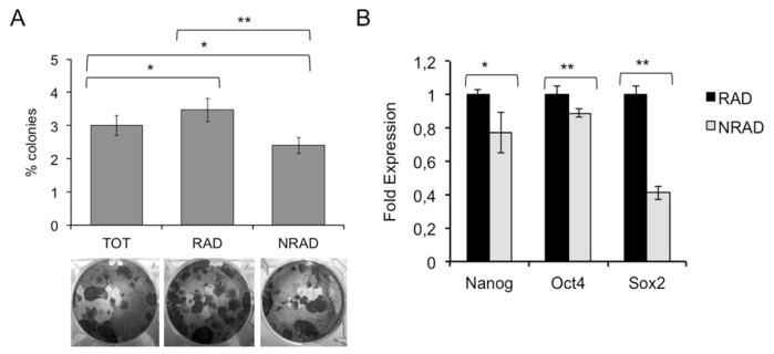
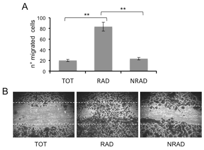
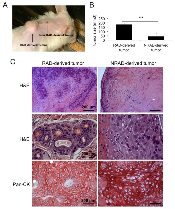
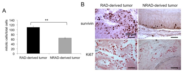
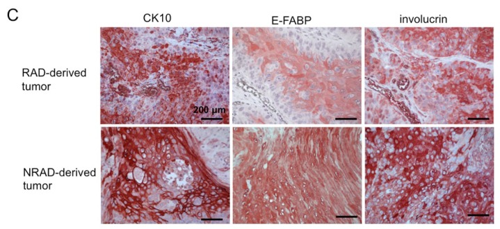
In human epidermis, keratinocyte stem cells (KSC) are characterized by high levels of β1-integrin, resulting in the rapid adhesion to type IV collagen. Since epithelial tumors originate from KSC, we evaluated the features of rapidly adhering (RAD) keratinocytes derived from primary human squamous cell carcinoma of the skin (cSCC). RAD cells expressed higher levels of survivin, a KSC marker, as compared to non-rapidly adhering (NRAD) cells. Moreover, RAD cells proliferated to a greater extent and were more efficient in forming colonies than NRAD cells. RAD cells also migrated significantly better than NRAD cells. When seeded in a silicone chamber and grafted onto the back skin of NOD SCID mice, RAD cells formed tumors 2-4 fold bigger than those derived from NRAD cells. In tumors derived from RAD cells, the mitotic index was significantly higher than in those derived from NRAD cells, while Ki-67 and survivin expression were more pronounced in RAD tumors. This study suggests that SCC RAD stem cells play a critical role in the formation and development of epithelial tumors.


Figures

Figure 1
Figure 1
β1-integrin levels in cSCC subpopulations. β1-integrin levels in RAD, NRAD and TOT cells were analyzed immediately after separation by Western blot. β-actin was used as loading control. Graph shows the average densitometry values normalized to actin, ** p < 0.01.
Figure 2
Figure 2
Proliferative ability of cSCCs subpopulations in vitro. (A) RAD, NRAD and TOT cells ability to proliferate in vitro was evaluated by CV staining; (B) RAD, NRAD and TOT cells were cultured for 72 h. BrdU incorporation was then evaluated by using FITC BrdU Flow Kit and analyzed by flow cytometry 72 h after the seeding. **p < 0.01; (C) Monoparametric histograms showing BrdU incorporation by FACS; (D) Density dot plots showing BrdU incorporation by FACS.
Figure 2
Figure 2
Proliferative ability of cSCCs subpopulations in vitro. (A) RAD, NRAD and TOT cells ability to proliferate in vitro was evaluated by CV staining; (B) RAD, NRAD and TOT cells were cultured for 72 h. BrdU incorporation was then evaluated by using FITC BrdU Flow Kit and analyzed by flow cytometry 72 h after the seeding. **p < 0.01; (C) Monoparametric histograms showing BrdU incorporation by FACS; (D) Density dot plots showing BrdU incorporation by FACS.
Figure 3
Figure 3
Expression of stem cell and differentiation markers in RAD, NRAD and TOT cells from cSCC. (A) Cells were analyzed immediately after separation, and levels of markers were determined by Western blot analysis. β-actin was used as loading control; (B) Bar graphs show the average densitometry values normalized to actin. *p < 0.05; **p < 0.01.
Figure 4
Figure 4
Analysis of stem-cell features in RAD and NRAD cells in vitro. (A) Clonal growth assessment of cSCC subpopulations by CFE. CFE was performed in triplicate in three independent experiments and quantification is shown in the upper panel. At the bottom, representative pictures of CFE obtained by growing cells at clonal density and stained with CV are shown; (B) mRNA expression of Nanog, Oct4 and Sox-2 by Real Time PCR. The different levels of gene expression in RAD vs. NRAD cells are shown. *p < 0.05; **p < 0.01.
Figure 5
Figure 5
Migration ability of cSCCs subpopulations. The migration of RAD, NRAD and TOT cells was determined by scratching assay. Cells were fixed and stained, and the number of migrated cells to the scratched surface area was counted. (A) Number of migrated cells expressed as the mean + SD from triplicate experiments, *p < 0.05, **p < 0.01; (B) Representative images of migrated cSCCs subpopulations in the scratching assay.
Figure 6
Figure 6
Tumorigenic ability of cSCC cells subpopulations in vivo. (A) Representative picture of RAD and NRAD-derived tumors formed by xenografting cSCC keratinocytes onto NOD/SCID mice; (B) Size of RAD and NRAD derived tumors as measured in three independent experiments. *p < 0.05, **p < 0.01; (C) Hematoxilin & Eosin and Pan-cytokeratin (CK) staining of RAD and NRAD-derived tumors. Scale bars = 200 μm.
Figure 7
Figure 7
RAD and NRAD-derived tumor characterization. (A) Mitotic Index representing the number of cells undergoing mitosis over total cells were counted in RAD and NRAD-derived tumors **p < 0.01; (B) Survivin and Ki67 expression in RAD and NRAD-derived tumors by immunohistochemistry. Scale bar = 200 μm; (C) K10, E-FABP and involucrin expression in RAD and NRAD-derived tumors by immunohistochemistry. Scale bar = 200 μm.
Figure 7
Figure 7
RAD and NRAD-derived tumor characterization. (A) Mitotic Index representing the number of cells undergoing mitosis over total cells were counted in RAD and NRAD-derived tumors **p < 0.01; (B) Survivin and Ki67 expression in RAD and NRAD-derived tumors by immunohistochemistry. Scale bar = 200 μm; (C) K10, E-FABP and involucrin expression in RAD and NRAD-derived tumors by immunohistochemistry. Scale bar = 200 μm.

References

    1. Lohmann C.M., Solomon A.R. Clinicopathologic variants of cutaneous squamous cell carcinoma. Adv. Anat. Pathol. 2001;8:27–36. - PubMed
    1. Morris R.J. Keratinocyte stem cells: targets for cutaneous carcinogens. J. Clin. Invest. 2000;106:3–8. - PMC - PubMed
    1. Lapouge G., Youssef K.K., Vokaer B., Achouri Y., Michaux C., Sotiropoulou P.A., Blanpain C. Identifying the cellular origin of squamous skin tumors. Proc. Natl. Acad. Sci. USA. 2011;108:7431–7436. - PMC - PubMed
    1. Jones P.H., Watt F.M. Separation of human epidermal stem cells from transit amplyfying cells on the basis of differences in integrin function and expression. Cell. 1993;73:713–724. - PubMed
    1. Tan D.W., Jensen K.B., Trotter M.W., Connelly J.T., Broad S., Watt F.M. Single-cell gene expression profiling reveals functional heterogeneity of undifferentiated human epidermal cells. Development. 2013;140:1433–1444. - PMC - PubMed

Publication types

MeSH terms