. 2013 Oct 29;128(18):2047-57.
doi: 10.1161/CIRCULATIONAHA.113.002887. Epub 2013 Sep 27.

Ten-eleven translocation-2 (TET2) is a master regulator of smooth muscle cell plasticity

Affiliations

Ten-eleven translocation-2 (TET2) is a master regulator of smooth muscle cell plasticity

Renjing Liu et al. Circulation. .

Abstract

Background: Smooth muscle cells (SMCs) are remarkably plastic. Their reversible differentiation is required for growth and wound healing but also contributes to pathologies such as atherosclerosis and restenosis. Although key regulators of the SMC phenotype, including myocardin (MYOCD) and KLF4, have been identified, a unifying epigenetic mechanism that confers reversible SMC differentiation has not been reported.

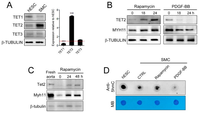
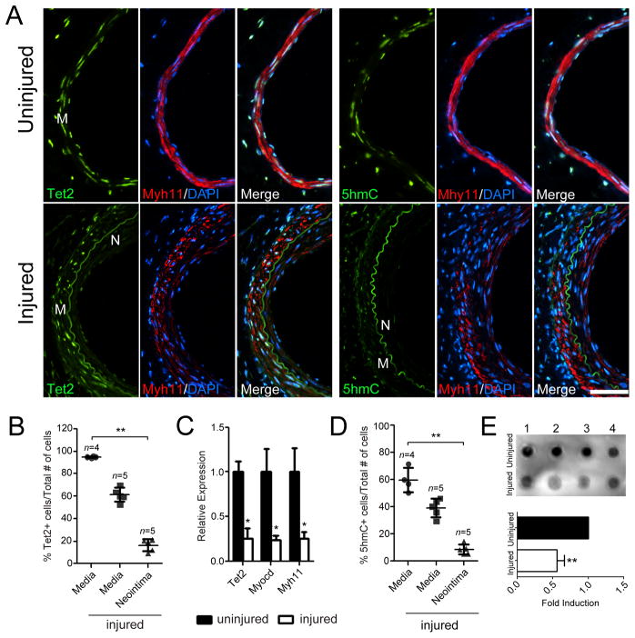
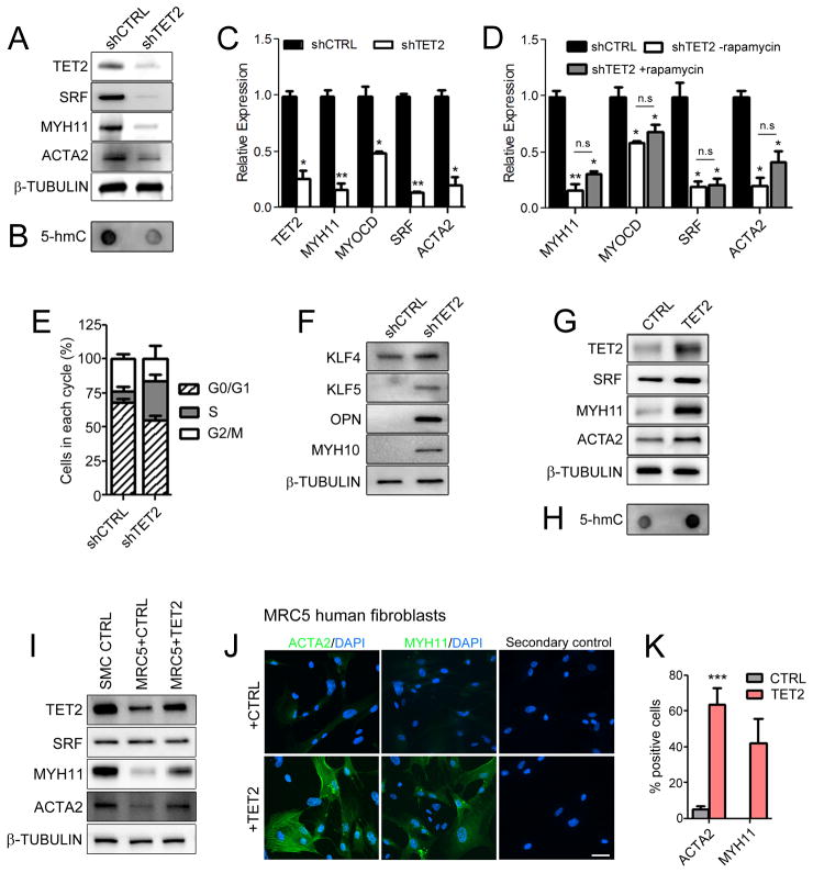
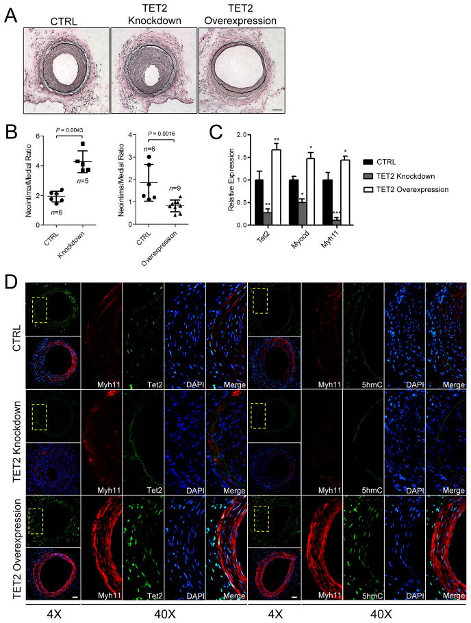
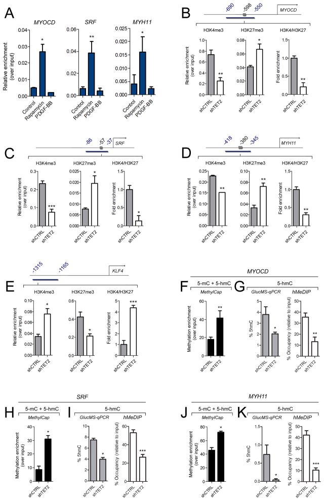
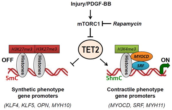
Methods and results: Using human SMCs, human arterial tissue, and mouse models, we report that SMC plasticity is governed by the DNA-modifying enzyme ten-eleven translocation-2 (TET2). TET2 and its product, 5-hydroxymethylcytosine (5-hmC), are enriched in contractile SMCs but reduced in dedifferentiated SMCs. TET2 knockdown inhibits expression of key procontractile genes, including MYOCD and SRF, with concomitant transcriptional upregulation of KLF4. TET2 knockdown prevents rapamycin-induced SMC differentiation, whereas TET2 overexpression is sufficient to induce a contractile phenotype. TET2 overexpression also induces SMC gene expression in fibroblasts. Chromatin immunoprecipitation demonstrates that TET2 coordinately regulates phenotypic modulation through opposing effects on chromatin accessibility at the promoters of procontractile versus dedifferentiation-associated genes. Notably, we find that TET2 binds and 5-hmC is enriched in CArG-rich regions of active SMC contractile promoters (MYOCD, SRF, and MYH11). Loss of TET2 and 5-hmC positively correlates with the degree of injury in murine models of vascular injury and human atherosclerotic disease. Importantly, localized TET2 knockdown exacerbates injury response, and local TET2 overexpression restores the 5-hmC epigenetic landscape and contractile gene expression and greatly attenuates intimal hyperplasia in vivo.

Conclusions: We identify TET2 as a novel and necessary master epigenetic regulator of SMC differentiation.

Keywords: cell differentiation; epigenomics; gene expression regulation; hyperplasia; muscle, smooth.


Conflict of interest statement

Conflict of Interest Disclosures: None.

Figures

Figure 1
Figure 1
TET2 is associated with the differentiated SMC state. (A) Western blot showing protein levels of TET1, TET2 and TET3 in human coronary artery smooth muscle cells (SMC) compared to human embryonic stem cells (hESC). Corresponding relative mRNA levels are shown on the right (qPCR). Relative expression of hESC is set to 1.0 and denoted by the red line. Data are presented as mean ± SD for three independent experiments. ***P<0.001. (B) Western blot showing TET2 and MYH11 levels in hCASMC following 50 nM rapamycin or 5 ng/ml PDGF-BB treatment. (C) Western blot comparing Tet2 and Mhy11 levels from freshly isolated mouse aorta tissue and cultured cells. (D) Dot blots for global 5-hmC expression using genomic DNA isolated from hESC and hCASMC treated as in (B) for 48 hours. Methylene blue (MB) staining demonstrates equal loading.
Figure 2
Figure 2
TET2 is downregulated during vascular injury. (A) TET2 and 5-hmC immunostaining of cross-sections from uninjured contralateral control mouse femoral arteries (top row) or injured femoral arteries (bottom row) collected 3 weeks following wire injury. Nuclei are stained with DAPI. (B) Quantification of TET2 positive cells divided by total number of DAPI positive cells in the uninjured (n=4) compared to injured (n=5) mouse femoral arteries. **P<0.01 relative to uninjured media. (C) qPCR for Tet2, Myocd and Myh11 gene expression in uninjured compared to injured femoral arteries 3 weeks post-injury. RNA was isolated from media and neointima from sections on slides using the Pinpoint isolation system. The adventitia was removed to ensure that only the media and neointima were used for analysis. *P<0.05 relative to uninjured arteries. (D) Quantification of 5-hmC positive cells divided by total number of DAPI positive cells in the uninjured (n=4) versus injured (n=5) femoral arteries. **P<0.01 relative to uninjured media. (E) Dot blot for global 5-hmC levels of uninjured and injured samples (n=4). Genomic DNA was isolated from tissue sections using the Pinpoint isolation system as in (C). Fold induction levels were calculated based on pixel intensity using Image Lab Software (**P<0.01). M = media, N = neointima. Scale bar, 50 μm. Error bars represent mean ± SD. n represent the number of individual mice. Two or more slides (12–18 sections) per animal were used for analysis.
Figure 3
Figure 3
TET2 and 5-hmC levels are high in mature human SMC and are lost in atherosclerotic lesions. (A) H&E staining (row 1), TET2 and MYH11 immunostaining (rows 2–5) of human coronary arteries with various degrees of atherosclerosis. The boxed areas in 4X sections are enlarged and shown in the lower panels. Scale bar, 50 μm. (B) Quantitation of the percent of TET2 expressing cells over total number of cells. n represent the number of individuals analyzed. Two sections from each patient (with five to eight random fields chosen) were captured and counted. Error bars represent mean ± SD. **P<0.01 relative to healthy subjects. (C) Dot blot of 5-hmC levels of the various human samples using the Pinpoint isolation system as in Figure 1E.
Figure 4
Figure 4
TET2 regulates SMC phenotype. (A) Western blot of contractile genes following TET2 knockdown (shTET2) in hCASMC. (B) Dot blot of 5-hmC in control (shCTRL) compared to shTET2 cells. (C–D) qPCR for contractile genes in shCTRL or shTET2 hCASMC treated with vehicle or 50 nM rapamycin for 24 hours. (E) Percentage of hCASMC in each cell cycle phase following TET2 knockdown compared to control. (F) Western blot for dedifferentiation genes in shTET2 cells compared to shCTRL cells. (G) Western blot of contractile markers in TET2 overexpressing hCASMC compared to control cells. (H) Dot blot of global 5-hmC in TET2 overexpressing hCASMC compared to control cells. (I) Western blot of SMC contractile markers in control and TET2 overexpressing MRC5 fibroblasts. (J) ACTA2 and MYH11 immunostaining of MRC5 fibroblasts infected with a control or TET2 overexpression virus. (K) Quantitation of ACTA2 and MYH11 positive cells in TET2 overexpressing MRC5 cells. Data are presented as mean ± SD for three independent experiments. *P<0.05, **P<0.01, ***P<0.001 compared control cells.
Figure 5
Figure 5
TET2 modulation alters 5-hmC levels and the intimal hyperplastic response to arterial injury. (A) EVG staining 3 weeks post-injury of femoral arteries transduced with either a control, TET2 knockdown or TET2 overexpressing virus. (B) Quantitation of the neointima/media ratio in each group. (C) RNA was isolated from the neointima of the control, TET2 knockdown or TET2 overexpression injured mice using the Pinpoint Isolation System and gene expression for Tet2, Myocd and Myh11 in each group was determined by qPCR. (D) Representative images of Myh11, Tet2 and 5-hmC immunostaining from each group. Boxed areas are enlarged on the right. Scale bar, 50 μm. n represent the number of individual mice analyzed. Two or more slides (12–18 sections) per animal were used for analysis. Data are presented as mean ± SD. *P<0.05, **P<0.01, ***P<0.001 compared to the control group.
Figure 6
Figure 6
TET2 binds to SMC promoters to regulate 5-hmC and modify histones at SMC loci. (A) ChIP assay demonstrating TET2 enrichment at SRF, MYOCD and MYH11 promoters during SMC differentiation. hCASMC were treated with rapamycin or PDGF-BB for 24 hours. Data are presented as mean relative enrichment over input ± SD of three biological repeats. *P<0.05, **P<0.01. (B–E) H3K4me3 and H3K27me3 ChIP-qPCR in shCTRL (gray bars) or shTET2 (white bars) hCASMC at various gene promoters. Primers were designed to encompass the CArG elements (denoted by the gray box). Primer locations are indicated in blue numbering, product is indicated as blue bar. Data are presented as mean relative enrichment over input ± SD of four biological replicates. *P<0.05, **P<0.01, ***P<0.001 compared to the shCTRL group. (F,H,J) DNA methylation as quantified by MethylCap. (G,I,K) 5-hMC levels as determined by GlucMS-qPCR or hMeDIP-qPCR. Data are presented as mean ± SD of four independent experiments. *P<0.05, **P<0.01, ***P<0.001 compared to the shCTRL group.
Figure 7
Figure 7
Proposed model of SMC phenotypic modulation control by TET2. Vascular injury releases a milieu of growth factors including PDGF-BB that can activate the mTORC1 pathway and promote SMC dedifferentiation to the synthetic phenotype. Rapamycin, an inhibitor of mTORC1, induces SMC differentiation. We now provide the first study to show that the mTORC1 pathway regulates SMC differentiation through regulation of TET2. In SMC, the balance between contractile and synthetic gene expression is governed by TET2, which results in changes in chromatin conformation and 5-hmC modifications at key promoters. An open chromatin conformation (as assessed by high levels of H3K4me3 and low expression of H3K27me3) as well as high levels of 5-hmC are seen at pro-contractile gene promoters in differentiated SMC, while TET2 promotes a closed chromatin formation at the KLF4 promoter.

Comment in

References

    1. Alexander MR, Owens GK. Epigenetic control of smooth muscle cell differentiation and phenotypic switching in vascular development and disease. Annu Rev Physiol. 2012;74:13–40. - PubMed
    1. Owens GK, Kumar MS, Wamhoff BR. Molecular regulation of vascular smooth muscle cell differentiation in development and disease. Physiol Rev. 2004;84:767–801. - PubMed
    1. Ito S, Shen L, Dai Q, Wu SC, Collins LB, Swenberg JA, He C, Zhang Y. Tet proteins can convert 5-methylcytosine to 5-formlcytosine and 5-carboxylcytosine. Science. 2011;333:1300–3. - PMC - PubMed
    1. Ito S, D’Alessio AC, Taranova OV, Hong K, Sowers LC, Zhang Y. Role of Tet proteins in 5mC to 5hmC conversion, ES-cell self-renewal and inner cell mass specification. Nature. 2010;466:1129–33. - PMC - PubMed
    1. Tahiliani M, Koh KP, Shen Y, Pastor WA, Bandukwala HS, Brudno Y, Agarwal S, Iyer LM, Liu DR, Aravind L, Rao A. Conversion of 5-methylcytosine to 5-hydroxymethylcytosine in mammalian DNA by MLL partner TET1. Science. 2009;324:930–5. - PMC - PubMed

Publication types

MeSH terms