Review
. 2013 Oct;93(4):382-96.
doi: 10.1007/s00223-013-9784-9.

A review of phosphate mineral nucleation in biology and geobiology

Review

A review of phosphate mineral nucleation in biology and geobiology

Sidney Omelon et al. Calcif Tissue Int. 2013 Oct.

Abstract

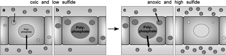
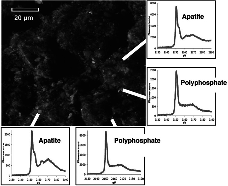
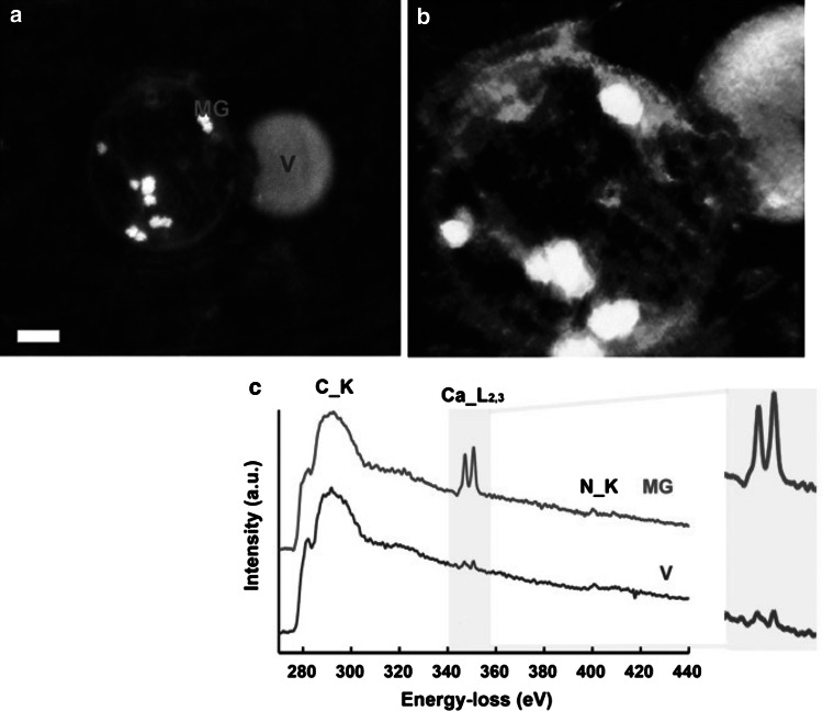
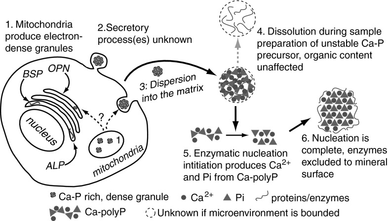
Relationships between geological phosphorite deposition and biological apatite nucleation have often been overlooked. However, similarities in biological apatite and phosphorite mineralogy suggest that their chemical formation mechanisms may be similar. This review serves to draw parallels between two newly described phosphorite mineralization processes, and proposes a similar novel mechanism for biologically controlled apatite mineral nucleation. This mechanism integrates polyphosphate biochemistry with crystal nucleation theory. Recently, the roles of polyphosphates in the nucleation of marine phosphorites were discovered. Marine bacteria and diatoms have been shown to store and concentrate inorganic phosphate (Pi) as amorphous, polyphosphate granules. Subsequent release of these P reserves into the local marine environment as Pi results in biologically induced phosphorite nucleation. Pi storage and release through an intracellular polyphosphate intermediate may also occur in mineralizing oral bacteria. Polyphosphates may be associated with biologically controlled apatite nucleation within vertebrates and invertebrates. Historically, biological apatite nucleation has been attributed to either a biochemical increase in local Pi concentration or matrix-mediated apatite nucleation control. This review proposes a mechanism that integrates both theories. Intracellular and extracellular amorphous granules, rich in both calcium and phosphorus, have been observed in apatite-biomineralizing vertebrates, protists, and atremate brachiopods. These granules may represent stores of calcium-polyphosphate. Not unlike phosphorite nucleation by bacteria and diatoms, polyphosphate depolymerization to Pi would be controlled by phosphatase activity. Enzymatic polyphosphate depolymerization would increase apatite saturation to the level required for mineral nucleation, while matrix proteins would simultaneously control the progression of new biological apatite formation.


Figures

Fig. 1
Fig. 1
Proposed phosphate uptake and release by Beggiatoa. a, b Under oxic conditions and exposure to low sulfide concentrations, phosphate is taken up by Beggiatoa and accumulated as polyphosphate. The phosphate concentration in the medium decreases. c, d When the conditions change to anoxia and exposure to sulfide increases, the Beggiatoa decompose polyphosphate and release phosphate. This leads to an increase in phosphate in the medium. Reprinted by permission from Macmillan Publishers Ltd: ISME J [89], copyright 2011
Fig. 2
Fig. 2
X-ray fluorescence micrograph and fluorescence spectra of phosphorus-rich regions in Effingham inlet sediment. Sedimentary phosphorus (red) appears as distinct, heterogeneously distributed submicrometer-sized particles against a comparatively uniform background of sedimentary aluminum (blue) and magnesium (green). On the basis of high-resolution X-ray spectroscopic characterization, about half of the 147 phosphorus-rich regions examined in our samples were found to be polyphosphate, whereas the other half were classified as apatite, a common calcium phosphate mineral. From Diaz et al. [80]. Reprinted with permission from American Association for the Advancement of Science (AAAS) (Color figure online)
Fig. 3
Fig. 3
Analytical electron microscopic evidence of vesicle–mitochondrial interactions in mineralizing osteoblasts. a High-angle annular dark-field scanning TEM image of a dense granule-containing mitochondrion associating with a vesicle within an osteoblast in a mineralized nodule. The sample was prepared by high-pressure freezing and freeze substitution (HPF-FS). (Scale bar = 200 nm). b Voltex projection of a 3D tomographic reconstruction showing a mitochondrion conjoined with a vesicle. Dense granules are evident within the mitochondrion. Sample was prepared by HPF-FS. c Electron energy loss spectroscopy (EELS) of specified areas within the mitochondrion and vesicle in a. The mitochondrial granule and vesicle show characteristic calcium L2 and L3 edges at 346 eV. All spectra display carbon edges. d Orthoslices at 10-nm intervals through the tomographic reconstruction showing the mitochondrion–vesicle interface. The mitochondrial membrane is discontinuous where it conjoins the vesicle (arrows) [131]
Fig. 4
Fig. 4
Proposed controlled apatite biomineralization schematic. (1) Mitochondria produce polyPs from phosphate sources that form complexes with calcium, producing discrete, electron-dense, Ca- and P-rich granules. (2) Granules may be processed through the trans-Golgi network (TGN), and then secreted via budding or exocytosis. If processed through the TGN, granules may associate with matrix and/or noncollagenous proteins as well as phosphatase enzymes. The secreted product is an amorphous Ca-/P-rich granule that contains matrix proteins. It is unknown if the granule is encapsulated. (3) The granule migrates to mineral nucleation sites within the collageneous matrix, where the noncollagenous proteins may play significant roles in granular interaction with the matrix. (4) During sample preparation, these unstable, amorphous granules may be artifactually dissolved so that only the remaining protein component is observed. These may be “crystal ghosts.” (5) If a phosphatase enzyme component of the unstable, amorphous precursor is activated within the matrix, the Ca–polyP component begins to transform into Ca2+ and Pi components. The local, high concentrations of Ca2+ and Pi nucleate apatite. As the apatite nucleus grows while the polyP depolymerizes, the protein component of the granule is excluded from the growing apatite crystal. This displaces the granule protein components to the surface. (6) The excluded proteins surround the apatite crystal surface, where they control crystal growth and shape, among other functions

References

    1. Piccoli PM, Candela PA. Apatite in igneous systems. Rev Mineral Geochem. 2002;48:255–292. doi: 10.2138/rmg.2002.48.6. - DOI
    1. Van Cappellen P, Berner RA. A mathematical model for the early diagenesis of phosphorus and fluorine in marine sediments; apatite precipitation. Am J Sci. 1988;288:289–333. doi: 10.2475/ajs.288.4.289. - DOI
    1. Thompson DW. On growth and form. New York: MacMillan; 1945.
    1. Quekett J. On the intimate structure of bone, as composing the skeleton, in the four great classes of animals, viz., mammals, birds, reptiles, and fishes, with some remarks on the great value of the knowledge of such structure in determining the affinities of minute fragments of organic remains. J Microsc. 1849;2:46–58.
    1. Crosby CH, Bailey J. The role of microbes in the formation of modern and ancient phosphatic mineral deposits. Front Microbiol. 2012;3:e241–e247. doi: 10.3389/fmicb.2012.00241. - DOI - PMC - PubMed

Publication types