. 2014 Feb;5(1):11-21.
doi: 10.1007/s12672-013-0160-z.

Aryl hydrocarbon receptor in breast cancer—a newly defined prognostic marker

Aryl hydrocarbon receptor in breast cancer—a newly defined prognostic marker

Ryoko Saito et al. Horm Cancer. 2014 Feb.

Abstract

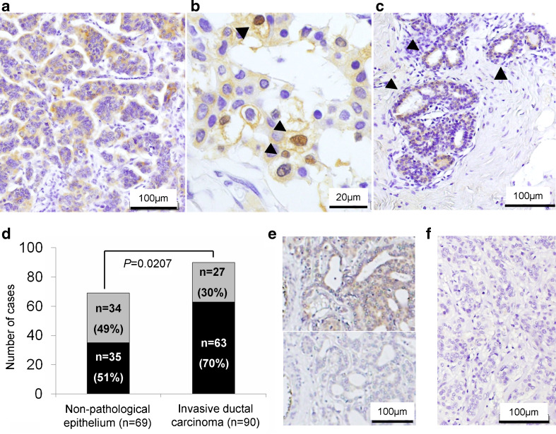
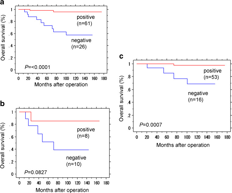
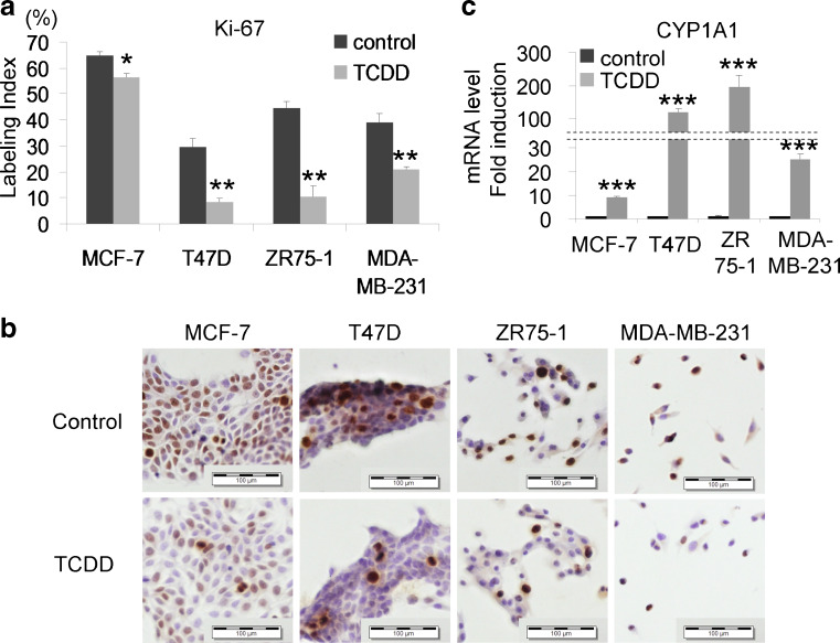
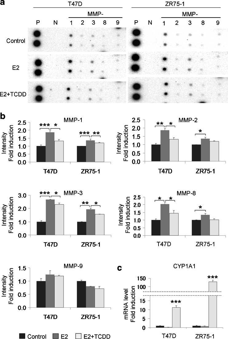
Aryl hydrocarbon receptor (AhR) has been reported to exert various anticancer effects upon breast carcinoma cells in vitro but its details have remained largely unknown. Therefore, we first examined the AhR status in 90 invasive ductal carcinoma patients using immunohistochemistry. We then performed in vitro studies including wound healing assay, invasion assay, and matrix metalloproteinase (MMP) protein array in order to further elucidate the roles of AhR signaling in breast carcinoma. The status of AhR immunoreactivity was inversely correlated with histological grade (P = 0.0135) and Ki-67 labeling index (LI; P = 0.0087) of the patients. In addition, results of both uni- and multivariate analyses revealed that AhR in carcinoma cells turned out an independent prognostic factor with a protective relative risk (P = 0.0179). An administration of 10 nM 2,3,7,8-tetrachlorodibenzo-p-dioxin (TCDD), a ligand of AhR, significantly decreased Ki-67 LI in an AhR-dependent fashion in MCF-7, T47D, ZR75-1, and MDA-MB-231. Wound healing and invasion assays performed in T47D and ZR75-1 further demonstrated that 10 nM TCDD inhibited estrogen-induced migration and invasion of cells. MMP proteins associated with AhR in breast carcinoma cells were also firstly identified. These results demonstrated that AhR in breast carcinoma cells is considered a newly defined histological prognostic parameter of the breast cancer patients and effects of AhR activation on proliferation and MMPs expression may be related to the relatively good clinical outcome of AhR-positive breast cancer patients.


Figures

Fig. 1
Fig. 1
ac Immunohistochemistry of AhR in human invasive ductal carcinoma (a, b) and nonpathological ductal epithelial cells (arrowhead; c). AhR immunoreactivity was mainly detected in the cytoplasm (a) and/or nuclei (arrowhead; b). d Black AhR positive, Gray AhR negative. The rate of AhR immunopositivity in carcinoma was significantly higher than that in nonpathological ductal epithelium (P = 0.0207). The statistical analysis was performed using χ 2 test. e The representative images of immunohistochemical absorption test using specimen of breast carcinoma and AhR antibody. Upper panel no AhR recombinant protein. Lower panel 10 μg/ml AhR recombinant protein. f The negative staining control using rabbit immunoglobulin fraction
Fig. 2
Fig. 2
Overall survival of 87 out of 90 cases with invasive ductal carcinoma according to AhR status of carcinoma cells (Kaplan–Meier method). a AhR immunoreactivity was significantly associated with better prognosis (P < 0.0001, logrank test). b, c Overall survival according to AhR with the ERα status separated. b In 18 ERα-negative cases, AhR immunoreactivity tend to be associated with better prognosis but the correlation did not reach the statistical significance (P = 0.0827). c In 69 ERα-positive cases, AhR immunoreactivity was significantly associated with better prognosis (P = 0.0007)
Fig. 3
Fig. 3
MCF-7, T47D, ZR75-1, and MDA-MB-231 were all treated with 10 nM TCDD for 24 h in phenol red- and FBS-free medium. a, b Ki-67 LI of both ER-positive and ER-negative breast cancer cell lines decreased by TCDD administration. Bars mean ± SE (n = 3). *P < 0.05, **P < 0.01 compared with control. c CYP1A1 was induced following TCDD administration, consistent with AhR activation by TCDD. Bars mean ± SE (n = 3). ***P < 0.001 compared with control. Scale bars 100 μm
Fig. 4
Fig. 4
a Wound healing assay in T47D and ZR75-1. T47D were exposed to 10 nM TCDD, 10 nM estrogen (E2) and/or 10 μM AhR antagonist (CH-223191), and ZR75-1 were exposed to 10 nM E2 and/or 10 nM TCDD in phenol red- and FBS-free medium for 24 h. Exposure to 10 nM E2 in serum-free medium for 24 h significantly induced migration of T47D and ZR75-1. A 10 nM TCDD administration significantly inhibited E2-dependent induction of cell migration in both cell lines. The inhibitory effects of TCDD on E2-dependent cell migration were significantly reduced by specific AhR antagonist in T47D cells. Bars mean ± SE (n = 4). *P < 0.05, **P < 0.01, ***P < 0.001. b We examined the effects of AhR on cell invasion using Matrigel-coated transwell chambers. T47D and ZR75-1 were treated with 10 nM E2 and/or 10 nM TCDD in phenol red- and FBS-free medium. After 24 h, the cells were plated in the upper chambers (5 × 105 cells/ml). Phenol red-free charcoal-stripped RPMI 1640 medium with 10 % FBS was used as a chemoattractant in the lower chamber. After 24 h, the cells invaded were counted. With 10 nM E2 administration, T47D and ZR75-1 invaded with forming larger clusters, and the number of invaded cells counted at three to five random fields in hotspot areas was significantly higher than that in control. A 10 nM TCDD administration significantly suppressed E2-dependent induction of cell invasion of T47D (P = 0.0431) and ZR75-1 (P = 0.0452). Bars mean ± SE (n = 3–5). *P < 0.05, **P < 0.01. c Cell proliferation assay using WST-8 method. Both T47D and ZR75-1 demonstrated no significant changes of the number of the cells between control and 10 nM E2 administration in serum-free medium for 24 h. Bars mean ± SE (n = 6). d We confirmed the activation of AhR by examining CYP1A1 induction. Bars mean ± SE (n = 3). ***P < 0.001 compared with control
Fig. 5
Fig. 5
a We examined MMPs protein expression and the effects of E2 and AhR on MMPs, using the human matrix metalloproteinase antibody array. b We examined the protein expression levels by measuring corrected relative immunointensity of each spots. Bars mean ± SE (n = 2). *P < 0.05, **P < 0.01, and ***P < 0.001. c We confirmed the activation of AhR by examining CYP1A1 induction. Bars mean ± SE (n = 3). ***P < 0.001 compared with control

Similar articles

Cited by

References

    1. Abdelrahim M, Smith R, 3rd, Safe S. Aryl hydrocarbon receptor gene silencing with small inhibitory RNA differentially modulates Ah-responsiveness in MCF-7 and HepG2 cancer cells. Mol Pharmacol. 2003;63(6):1373–1381. doi: 10.1124/mol.63.6.1373. - DOI - PubMed
    1. Adachi J, Mori Y, Matsui S, Takigami H, Fujino J, Kitagawa H, Miller CA, 3rd, Kato T, Saeki K, Matsuda T. Indirubin and indigo are potent aryl hydrocarbon receptor ligands present in human urine. J Biol Chem. 2001;276(34):31475–31478. doi: 10.1074/jbc.C100238200. - DOI - PubMed
    1. Beischlag TV, Luis Morales J, Hollingshead BD, Perdew GH. The aryl hydrocarbon receptor complex and the control of gene expression. Crit Rev Eukaryot Gene Expr. 2008;18(3):207–250. doi: 10.1615/CritRevEukarGeneExpr.v18.i3.20. - DOI - PMC - PubMed
    1. Bertazzi PA, Zocchetti C, Guercilena S, Consonni D, Tironi A, Landi MT, Pesatori AC. Dioxin exposure and cancer risk: a 15-year mortality study after the “Seveso accident”. Epidemiology. 1997;8(6):646–652. - PubMed
    1. Caldarella A, Crocetti E, Bianchi S, Vezzosi V, Urso C, Biancalani M, Zappa M. Female breast cancer status according to ER, PR and HER2 expression: a population based analysis. Pathol Oncol Res. 2011;17(3):753–758. doi: 10.1007/s12253-011-9381-z. - DOI - PubMed

MeSH terms