. 2013 Nov 15;288(46):33027-36.
doi: 10.1074/jbc.M113.517540. Epub 2013 Sep 27.

Sequential proteolytic processing of the triggering receptor expressed on myeloid cells-2 (TREM2) protein by ectodomain shedding and γ-secretase-dependent intramembranous cleavage

Affiliations

Sequential proteolytic processing of the triggering receptor expressed on myeloid cells-2 (TREM2) protein by ectodomain shedding and γ-secretase-dependent intramembranous cleavage

Patrick Wunderlich et al. J Biol Chem. .

Abstract

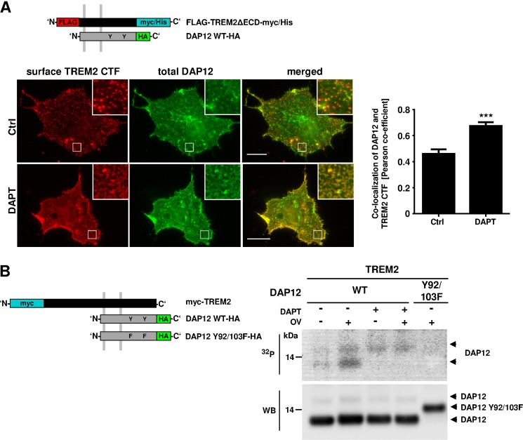
Triggering receptor expressed on myeloid cells-2 (TREM2) and its signaling adaptor protein TYROBP/DAP12 play important roles in signal transduction in dendritic cells, osteoclasts, tissue macrophages, and microglia. Recently, TREM2 variants have been shown to be linked to late onset Alzheimer disease. Here, we demonstrate that TREM2 undergoes sequential proteolytic processing by ectodomain shedding and intramembrane proteolysis. The C-terminal fragment (CTF) of TREM2 generated by ectodomain shedding is cleaved by γ-secretase. Importantly, pharmacologic and genetic γ-secretase inhibition resulted in accumulation of TREM2 CTF at the plasma membrane that also interacts with the signaling adaptor protein DAP12. Thus, the accumulated TREM2 CTF thereby might limit the interaction of DAP12 with the functional full-length receptor, resulting in decreased DAP12 phosphorylation and impaired metabolism of phosphatidylinositol 4,5-bisphosphate. Together, these data demonstrate γ-secretase-mediated intramembranous proteolysis of TREM2 and functionally link two Alzheimer disease-associated proteins in one signaling pathway.

Keywords: Alzheimer Disease; Intramembrane Proteolysis; Membrane; Presenilin; Protein Phosphorylation; Secretases; Signaling.


Figures

FIGURE 1.
FIGURE 1.
Shedding of the TREM2 ectodomain. A, COS7 cells were co-transfected with DAP12-HA and myc-TREM2-GFP (scheme). After 24 h, cells were treated with 10 μm batimastat or dimethyl sulfoxide as solvent control for 1 h. Surface localization of TREM2 was visualized by surface staining using anti-myc primary and Alexa Fluor 594-coupled secondary antibodies (see “Experimental Procedures”). Scale bars represent 20 μm. Bar chart shows quantification of TREM2 signals in randomly chosen areas of 75 × 75 pixels (Ctrl, n = 25; batimastat, n = 40 areas). B, HEK293 cells co-transfected with FLAG-DAP12-HA and myc-TREM2-GFP were incubated in the presence or absence of batimastat (10 μm) for 24 h in serum-free medium. Cellular membranes were isolated and conditioned medium precipitated with TCA. Proteins were separated by SDS-PAGE, and TREM2 was detected by Western immunoblotting with anti-GFP (membrane) or anti-myc antibodies (medium). The different bands detected for TREM2 in membrane fractions and for the secreted TREM2 ECD in medium might represent different glycosylation state variants. Bar charts show the quantification of TREM2 ECD/FL ratios (left panel) and TREM2 CTF/FL ratios (right panel) (n = 3). Statistical analysis was done by two-tailed t test. Error bars, S.E. **, p < 0.01; ***, p < 0.001.
FIGURE 2.
FIGURE 2.
Cleavage of TREM2 C-terminal fragments by γ-secretase. A–C, native HEK293 cells (A) or HEK293 cells stably overexpressing PS1 WT or PS1 DN variants (B and C) were co-transfected with FLAG-DAP12-HA and FLAG-TREM2-myc/His (A and B) or with FLAG-TREM2ΔECD-myc/His (C) and cultured for 24 h. Membrane proteins were separated by SDS-PAGE, and TREM2 variants were detected by Western immunoblotting with anti-myc antibody. Bar charts show the quantification of TREM2 CTF/FL ratios (A and B; n = 3) or relative levels of TREM2 CTFs (C; n = 2). Statistical determination was done by a two-tailed t test (A) (n = 3) or one-way analysis of variance ANOVA (B) ( n = 3). D, in vitro γ-secretase activity assay. Membranes of HEK293 cells expressing myc-TREM2-GFP were isolated and incubated for 2 h in the absence or presence of 10 μm DAPT. Reaction mixtures were separated by centrifugation into membrane (Membr.) and supernatant (Sup.) fractions. TREM2 CTF-GFP in the membrane fraction and soluble TREM2 ICD-GFP in the supernatant was detected by Western immunoblotting with anti-GFP antibodies. The soluble TREM2 ICD-GFP was strongly reduced after incubation with DAPT. Bar chart shows the quantification of TREM2 ICD/CTF ratios (n = 2). Error bars, S.E. **, p < 0.01; ***, p < 0.001.
FIGURE 3.
FIGURE 3.
TREM2 CTFs accumulate at the cell surface after γ-secretase inhibition. A, HEK293 cells were transfected with FLAG-TREM2-myc/His in combination with FLAG-DAP12-HA and incubated for 24 h in absence or presence of 10 μm DAPT. Surface proteins were labeled with sulfo-NHS-biotin for 30 min. Cells were lysed and biotin-labeled proteins precipitated with streptavidin-coated agarose beads. Aliquots of cell lysates and streptavidin-precipitates were separated by SDS-PAGE, and TREM2 was detected by Western immunoblotting with anti-myc antibodies. Treatment with DAPT increases the cell surface expression of TREM2. Bar graphs show the quantification of TREM2 CTF/FL ratios (n = 3). B, COS7 cells were co-transfected with DAP12-HA and FLAG-TREM2ΔECD-myc/His and incubated for 24 h in presence or absence of 10 μm DAPT. TREM2 CTF was visualized at the cell surface of living cells by anti-FLAG primary and Alexa Fluor 488-coupled secondary antibodies. Bar graph shows the quantification of TREM2 CTF-positive structures in five randomly chosen areas of 75 × 75 pixels in 10 cells each. Insets show enlarged images of boxed areas. Scale bars represent 20 μm. Statistical analyses were done by using a two-tailed t test. Error bars, S.E. *, p < 0.05; ***, p < 0.001.
FIGURE 4.
FIGURE 4.
Inhibition of γ-secretase alters the interaction of TREM2 with DAP12 and impairs DAP12 phosphorylation. A, COS7 cells expressing DAP12-HA and FLAG-TREM2ΔECD-myc/His (scheme) were incubated in the presence or absence of 10 μm DAPT for 24 h. Surface TREM2 CTF was visualized using anti-FLAG primary and Alexa Fluor 594-coupled secondary antibodies. After the specific cell surface staining, cells were fixed and permeabilized, and total DAP12 was stained using anti-HA primary and Alexa Fluor 488-coupled secondary antibodies. Co-localization of DAP12 and TREM2 CTF was analyzed by the Pearson co-efficient in four randomly chosen areas of four different cells. Insets show enlarged images of boxed areas. Scale bars represent 20 μm. Statistical analysis was done by two-tailed t test (p < 0.001). Error bars, S.E. ***, p < 0.001. B, HEK293 cells co-expressing myc-TREM2 and DAP12-HA were incubated in presence or absence of 10 μm DAPT for 24 h. The DAP12 Y92F/Y103F-HA variant lacking potential phosphorylation sites in the ITAM domain was co-transfected with TREM2 as a control (last lane). Cells were starved for 30 min in phosphate-free medium and then incubated with 32Pi for 1 h. TREM2 was activated by incubation with 10 μg/ml anti-myc antibody. After further incubation for 1 h in the presence or absence of 200 μm orthovanadate (OV), cells were lysed, and DAP12 was precipitated with anti-HA antibodies. Proteins were separated on a Tris-Tricine gel and transferred onto a nitrocellulose membrane. Radiolabeled proteins were visualized by autoradiography (upper panel, 32P). After exposure, DAP12-HA was detected on the same membrane by Western immunoblotting with anti-HA antibodies (lower panel, WB).
FIGURE 5.
FIGURE 5.
Impaired DAP12 signaling upon γ-secretase inhibition affects PIP2 metabolism. A, COS7 cells expressing the PIP2 sensor PH-PLCδ-GFP in combination with myc-TREM2-mCherry and DAP12-HA (scheme) were incubated for 24 h in presence or absence of 10 μm DAPT. Fluorescence of the PIP2 sensor was measured every 10 min for 120 min. c-myc antibodies (10 μg/ml) were applied after 20 min (dashed line). The graph shows the quantification of fluorescence intensities plotted as ΔF/F0 against time for cells incubated with (blue line) or without DAPT (red line). The fluorescence in cells expressing the PIP2 sensor alone served as control (gray line). Statistical determination was done by two-way analysis of variance (+DAPT, n = 4; −DAPT, n = 6). Error bars, S.E. **, p < 0.01; ***, p < 0.001. ns, not significant. B, schematic drawing shows the proposed role of TREM2 processing in membrane-proximal signaling with either active (upper panel) or inhibited γ-secretase (lower panel). In absence of ligand (upper panel, left), the ectodomain of membrane-associated TREM2 FL is cleaved by a sheddase. The resultant TREM2 CTF is then cleaved by γ-secretase. In the presence of ligand, TREM2 signals to DAP12 (upper panel, right). Upon inhibition of γ-secretase, TREM2 CTFs accumulate after ectodomain shedding (lower panel, left). Accumulated CTFs trap DAP12 co-receptors, thereby preventing the interaction with full-length TREM2 receptors (lower panel, right; see “Discussion” for details). L, ligand; γ, γ-secretase; P, phosphate.

References

    1. Colonna M. (2003) TREMs in the immune system and beyond. Nat. Rev. Immunol. 3, 445–453 - PubMed
    1. Bouchon A., Dietrich J., Colonna M. (2000) Cutting edge: inflammatory responses can be triggered by TREM-1, a novel receptor expressed on neutrophils and monocytes. J. Immunol. 164, 4991–4995 - PubMed
    1. Bouchon A., Hernández-Munain C., Cella M., Colonna M. (2001) A DAP12-mediated pathway regulates expression of CC chemokine receptor 7 and maturation of human dendritic cells. J. Exp. Med. 194, 1111–1122 - PMC - PubMed
    1. Sessa G., Podini P., Mariani M., Meroni A., Spreafico R., Sinigaglia F., Colonna M., Panina P., Meldolesi J. (2004) Distribution and signalling of TREM2/DAP12, the receptor system mutated in the human PLOSL dementia. Eur. J. Neurosci. 20, 2617–2628 - PubMed
    1. Prada I., Ongania G. N., Buonsanti C., Panina-Bordignon P., Meldolesi J. (2006) Triggering receptor expressed in myeloid cells 2 (TREM2) trafficking in microglial cells: continuous shuttling to and from the plasma membrane regulated by cell stimulation. Neuroscience 140, 1139–1148 - PubMed

Publication types

MeSH terms

Substances