Review
. 2014 Jan;99(1):16-34.
doi: 10.1113/expphysiol.2013.071951. Epub 2013 Sep 27.

Purinergic signalling: from discovery to current developments

Affiliations
Free PMC article
Review

Purinergic signalling: from discovery to current developments

Geoffrey Burnstock. Exp Physiol. 2014 Jan.
Free PMC article

Abstract

This lecture is about the history of the purinergic signalling concept. It begins with reference to the paper by Paton & Vane published in 1963, which identified non-cholinergic relaxation in response to vagal nerve stimulation in several species, although they suggested that it might be due to sympathetic adrenergic nerves in the vagal nerve trunk. Using the sucrose gap technique for simultaneous mechanical and electrical recordings in smooth muscle (developed while in Feldberg's department in the National Institute for Medical Research) of the guinea-pig taenia coli preparation (learned when working in Edith Bülbring's smooth muscle laboratory in Oxford Pharmacology), we showed that the hyperpolarizations recorded in the presence of antagonists to the classical autonomic neurotransmitters, acetylcholine and noradrenaline, were inhibitory junction potentials in response to non-adrenergic, non-cholinergic neurotransmission, mediated by intrinsic enteric nerves controlled by vagal and sacral parasympathetic nerves. We then showed that ATP satisfied the criteria needed to identify a neurotransmitter released by these nerves. Subsequently, it was shown that ATP is a cotransmitter in all nerves in the peripheral and central nervous systems. The receptors for purines and pyrimidines were cloned and characterized in the early 1990 s, and immunostaining showed that most non-neuronal cells as well as nerve cells expressed these receptors. The physiology and pathophysiology of purinergic signalling is discussed.


Figures

Figure 1
Figure 1. Changes in membrane potential and mechanical response recorded with a sucrose gap method
A, hyperpolarizations recorded in smooth muscle of the atropinized guinea-pig taenia coli in response to transmural stimulation of the intramural nerves remaining after degeneration of the adrenergic nerves by treatment of the animal with 6-hydroxydopamine (250 mg kg−1i.p. for 2 successive days) 7 days previously. Upper trace shows responses to low-frequency stimulation (1 s−1). Note the individual hyperpolarizations and rebound excitation (spike and contraction) following cessation of stimulation. Lower trace shows stimulation at higher frequencies, illustrating summed hyperpolarization and relaxation. [Reproduced from Burnstock (1972) with permission from the American Society for Pharmacology and Experimental Therapeutics.]B, transmural field stimulation (0.5 ms, 0.033 Hz, 8 V) of the taenia coli evoked transient hyperpolarizations in the presence of atropine (0.3 μm) and guanethidine (4 μm). Tetrodotoxin (TTX; 3 μm) added to the superfusing Krebs solution (applied at arrow) rapidly abolished the response to transmural field stimulation, establishing that the hyperpolarizations were inhibitory junction potentials in response to non-adrenergic, non-cholinergic (NANC) neurotransmission. [Reproduced from Burnstock (1986), reproduced with kind permission of Blackwell Publishing.]
Figure 2
Figure 2. Photographs of Albert Szent-Györgyi and Pamela Holton
Figure 3
Figure 3. Evidence that ATP satisfied the criteria for its establishment as a neurotransmitter in the guinea-pig taenia coli and urinary bladder
A, left-hand side shows responses of the guinea-pig taenia coli to NANC inhibitory nerve stimulation (NS; 1 Hz, 0.5 ms pulse duration, for 10 s at supramaximal voltage) mimicked by ATP (2 × 10⁻⁶ m). Atropine (1.5 × 10⁻⁷ m), guanethidine (5 × 10⁻⁶ m) and sodium nitrite (7.2 × 10⁻⁴ m) were present. [From Burnstock & Wong (1978), reproduced with kind permission of the Nature Publishing Group.]A, right-hand side shows a comparison of the NANC contractile responses of the guinea-pig bladder strip to intramural nerve stimulation (NS; 5 Hz, 0.2 ms pulse duration and supramaximal voltage) mimicked by exogenous ATP (8.5 μm). Atropine (1.4 μm) and guanethidine (3.4 μm) were present throughout. [From Burnstock et al. (1978), reproduced with kind permission of the Nature Publishing Group.]B, effect of changing the calcium ion (Ca2+) concentration on the release of ATP (measured with the firefly luciferin/luciferase technique) from the guinea-pig isolated bladder strip during stimulation of NANC nerves. Upper trace is a mechanical recording of changes in tension (in grams) during intramural nerve stimulation (NS; 20 Hz, 0.2 ms pulse duration, supramaximal voltage for 20 s). Lower histograms show the concentration of ATP in consecutive 20 s fractions of the superfusate. The Ca2+ concentration in the superfusate varied as follows: 2.5 (normal Krebs; a) 0.5 (b), 0.25 (c) and 2.5 mm (d). The successive contractions were separated by 60 min intervals as indicated by the breaks in the mechanical trace. Atropine (1.4 μm) and guanethidine (3.4 μm) were present throughout. [From Burnstock et al. (1978), reproduced with kind permission of the Nature Publishing Group.]C, the effect of α,β-methylene ATP (α,β-meATP) receptor desensitization on the responses to nerve stimulation (↑), ATP (open triangles) and histamine (Hist). Atropine (1 μm) and guanethidine (3.4 μm) were present throughout. Top panel shows control responses. Bottom panel shows that α,β-me ATP desensitization, reached by five successive applications (filled triangles) at approximately 4 min intervals, completely abolished nerve-mediated (↑) and ATP-induced responses (open triangles); However, histamine-induced contraction was only slightly reduced. [Reproduced from Kasakov & Burnstock (1983), with permission.]
Figure 4
Figure 4. Purinergic neuromuscular transmission depicting the synthesis, storage, release and inactivation of ATP
Adenosine triphosphate, stored in vesicles in nerve varicosities, is released by exocytosis to act on postjunctional receptors for ATP on smooth muscle. The ATP is broken down extracellularly by ATPases and 5′-nucleotidase to adenosine, which is taken up by varicosities to be resynthesized and reincorporated into vesicles. Adenosine is broken down further by adenosine deaminase (A. deaminase) to inosine and hypoxanthine and removed by the circulation. [From Burnstock (1972), reproduced with permission from the American Society for Pharmacology and Experimental Therapeutics.]
Figure 5
Figure 5. Evidence for ATP as a cotransmitter with noradrenaline in sympathetic nerves supplying the guinea-pig vas deferens
A, excitatory junction potentials (EJPs) in response to repetitive stimulation of sympathetic nerves (white dots) in the guinea-pig vas deferens. The upper trace records the tension, the lower trace the electrical activity of the muscle recorded extracellularly by the sucrose gap method. Note both summation and facilitation of successive junction potentials. At a critical depolarization threshold, an action potential is initiated, which results in contraction. [From Burnstock & Costa (1975), reproduced with permission of Chapman and Hall.]B, the effect of α,β-methylene ATP (α,β-meATP) on EJPs recorded from the guinea-pig vas deferens (intracellular recording). The control responses to stimulation of the motor nerves at 0.5 Hz are shown on the left. After at least 10 min in the continuous presence of α,β-meATP, EJPs were recorded using the same stimulation parameters. The EJPs were abolished in the presence of α,β-meATP (3 × 10⁻⁶m). [Reproduced from Sneddon & Burnstock (1984), with permission of Elsevier.]C, spritzed ATP, but not noradrenaline (NA), mimicked the EJP recorded in the vas deferens [Reproduced from Burnstock & Verkhratsky (2012), with permission of Springer.]
Figure 6
Figure 6. Schematic diagram of sympathetic cotransmission
Adenosine triphosphate released from small agranular vesicles and noradrenaline (NA) released from small granular vesicles (SGV) act on P2X and α1-adrenoceptors on smooth muscle, respectively. ATP acting on inotropic P2X receptors evokes excitatory junction potentials (EJPs), increase in [Ca2+]i and fast contraction, while occupation of metabotropic α1-adrenoceptors leads to production of inositol trisphosphate (InsP3), increase in [Ca2+]i and slow contraction. Neuropeptide Y (NPY) stored in large granular vesicles (LGV) acts on release both as a prejunctional inhibitory modulator of release of ATP and NA and as a postjunctional modulatory potentiator of the actions of ATP and NA. Nucleotidases are released from nerve varicosities, and are also present as ectonucleotidases to break ATP down to adenosine (ADO), which acts as a prejunctional modulator of ATP and NA release via A1 receptors. Noradrenaline is also a prejunctional modulator via α2-adrenoceptors. [Modified from Burnstock (2009c), and reproduced with permission from Elsevier.]
Figure 7
Figure 7. Structure of P2X receptors with respect to that of other channels
A, channel subunits that have two transmembrane domains. B, nicotinic and glutamate receptor families. [Reproduced from North (1996), with permission from Elsevier.]
Figure 8
Figure 8. The architecture of P2X receptors
Stereoview of the homotrimeric P2X4 structure viewed parallel to the membrane. Each subunit is depicted in a different colour. N-acetyl-d-glucosamine and glycosylated asparagine residues are shown in stick representation. The grey bars suggest the boundaries of the outer (out) and inner leaflets (in) of the membrane bilayer. [Reproduced from Kawate et al. (2009), with permission from the Nature Publishing Group.]
Figure 9
Figure 9. A schematic representation of short-term purinergic signalling, showing the interactions of ATP released from perivascular nerves and from the endothelium (Endoth.) controlling vascular tone
Adenosine triphosphate is released from endothelial cells during hypoxia to act mainly on endothelial P2Y receptors, leading to the production of endothelium-derived relaxing factor (EDRF; nitric oxide) and subsequent vasodilatation (−). In contrast, ATP released as a cotransmitter with noradrenaline (NA) from perivascular sympathetic nerves at the adventitia (Advent.)–muscle border produces vasoconstriction (+) via P2X receptors on the muscle cells. Adenosine (ADO), resulting from breakdown of ATP by ectoenzymes, later produces vasodilatation by direct action on the muscle via P1 receptors and acts on the perivascular nerve terminal varicosities to inhibit transmitter release. [From Burnstock (1987), reproduced with permission from S. Karger AG, Basel.]
Figure 10
Figure 10. Schematic overview of purinergic signalling mechanisms that regulate long-term, trophic effects
Extracellular nucleotides and nucleosides bind to purinergic receptors coupled to signal-transducing effector molecules. Activation of the effectors leads to generation of second messengers and/or stimulation of protein kinases that regulate expression of genes needed for long-term, trophic actions. In some cases, P2X receptors, such as P2X7, are also coupled to protein kinase cascades and can mediate proliferation and apoptosis. Cell-specific and/or receptor subtype-specific differences are likely to account for variations in signalling pathways and functional outcomes. It should be noted that the list of elements is not meant to be all-inclusive. Other protein kinases, e.g. MEK, PI3K, are upstream of the listed kinases involved in purinergic signalling, while others are downstream, e.g. p70S6K. In addition, dashed arrows indicate that not all listed elements are activated by the upstream component, e.g. not all P1 receptors are coupled to all listed effectors. Abbreviations: AC, adenylyl cyclase; AP-1, activator protein-1; CaMK, calcium–calmodulin protein kinase; CREB, cyclic AMP response element binding protein; DG, diacylglycerol; GSK, glycogen synthase kinase; IP3, inositol trisphosphate; MAPKs, mitogen-activated protein kinases [including extracellular signal-regulated protein kinase (ERK), p38 MAPK and stress-activated protein kinase (SAPK)/c-Jun N-terminal kinase (JNK)]; MEK, MAPK/ERK kinase; NO, nitric oxide; PG, prostaglandin; PI3K, phosphoinositide 3-kinase; PI-PLC, phosphatidylinositol-specific phospholipase C; PKA, protein kinase A; PKC, protein kinase C; PLA, phospholipase A; PLD, phospholipase D; and STAT3, signal transducer and activator of transcription-3. [Modified from Burnstock (2007b), with permission from the American Physiological Society.]
Figure 11
Figure 11. Predicted membrane topography of ectonucleotidases, consisting of the ectonucleoside triphosphate diphosphohydrolase (E-NTPDase) family (CD39), the ecto-nucleotide pyrophosphatase/phosphodiesterase family, alkaline phosphatases and ecto-5′-nucleotidase (CD73)
[Modified from Zimmermann (2001), with permission from Wiley-Liss, Inc.].
Figure 12
Figure 12. Three P2 receptor subtypes, P2X1, P2Y1 and P2Y12, are involved in ADP-induced platelet activation
Clopidogrel is a P2Y12 receptor blocker that inhibits platelet aggregation and is in highly successful use for the treatment of thrombosis and stroke. A P2Y1 receptor antagonist, MRS 2500, inhibits shape change. [Modified from Kunapuli & Daniel (1998), with permission from Portland Press Ltd.]
Figure 13
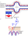
Figure 13. Purinergic mechanosensory transduction
A, schematic representation of the hypothesis for purinergic mechanosensory transduction in tubes (e.g. ureter, vagina, salivary duct, bile duct and gut) and sacs (e.g. urinary bladder, gall bladder and lung). It is proposed that distension leads to release of ATP from epithelium lining the tube or sac, which then acts on P2X3 and/or P2X2/3 receptors on subepithelial sensory nerves to convey sensory/nociceptive information to the CNS. [From Burnstock (1999), reproduced with permission from Blackwell Publishing.]B, schematic diagram of a novel hypothesis about purinergic mechanosensory transduction in the gut. It is proposed that ATP released from mucosal epithelial cells during moderate distension acts preferentially on P2X3 and/or P2X2/3 receptors on low-threshold subepithelial intrinsic sensory nerve fibres (labelled with calbindin) to modulate enteric reflexes. The ATP released during extreme (colic) distension also acts on P2X3 and/or P2X2/3 receptors on high-threshold extrinsic sensory nerve fibres [labelled with isolectin B4 (IB4)] that send messages via the dorsal root ganglia (DRG) to pain centres in the CNS. [From Burnstock (2001), reproduced with permission from Wiley.]
Figure 14
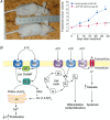
Figure 14. Reduction of cancer cell growth by nucleotides
A, left-hand panel shows the effect of ATP on the growth of implanted hormone refractory prostate cancer DU145 tumour cells in vivo after 60 days initial growth; the lower mouse received ATP treatment versus no treatment in the upper mouse. A, right-hand panel shows the effect of ATP on the fractional growth of DU145 tumour cells in vivo after 60 days initial growth. [Reproduced from Shabbir et al. (2008), with permission from Blackwell Publishing.]B, different mechanisms by which P2 receptor subtypes might alter cancer cell function. The P2Y1 and P2Y2 receptors affect the rate of cell proliferation by modulating adenylyl cyclase (AC) and altering the intracellular levels of cAMP, or by increasing the intracellular level of Ca2+ through the phospholipase C (PLC) pathway. P2X5 and P2Y11 receptor activation mediates differentiation, which switches the cell cycle to antiproliferation. The P2X7 receptor activates the apoptotic cell death caspase enzyme system. Abbreviations: DAG, diacylglycerol; Ins(1,4,5)P3, inositol (1,4,5)-trisphosphate; PtdIns(4,5)P2, phosphatidylinositol (4,5)-bisphosphate. (Reproduced from White & Burnstock, , with permission from Elsevier.)
Figure 15
Figure 15. Graph showing the number of papers published on P2 purinergic signalling between 1972 and the end of 2012

References

    1. Abbracchio MP. Burnstock G. Purinoceptors: are there families of P2X and P2Y purinoceptors. Pharmacol Ther. 1994;64:445–475. - PubMed
    1. Abbracchio MP. Burnstock G. Purinergic signalling: pathophysiological roles. Jpn J Pharmacol. 1998;78:113–145. - PubMed
    1. Agboh KC, Webb TE, Evans RJ. Ennion SJ. Functional characterization of a P2X receptor from Schistosoma mansoni. J Biol Chem. 2004;279:41650–41657. - PubMed
    1. Abbracchio MP, Burnstock G, Verkhratsky A. Zimmermann H. Purinergic signalling in the nervous system: an overview. Trends Neurosci. 2009;32:19–29. - PubMed
    1. Bailey MA, Shirley DG, King BF, Burnstock G, Unwin RJ. Extracellular nucleotides and renal function. In: Alpern RJ, Herbert SCG, editors. The Kidney: Physiology and Pathophysiology. San Diego: Elsevier; 2007. pp. 425–443.

MeSH terms