. 2013 Nov 1;191(9):4665-75.
doi: 10.4049/jimmunol.1300581. Epub 2013 Sep 27.

Identification of a tissue-specific, C/EBPβ-dependent pathway of differentiation for murine peritoneal macrophages

Affiliations

Identification of a tissue-specific, C/EBPβ-dependent pathway of differentiation for murine peritoneal macrophages

Derek W Cain et al. J Immunol. .

Abstract

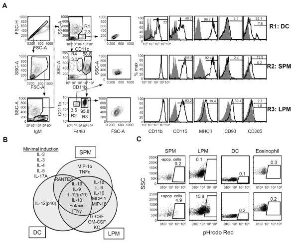
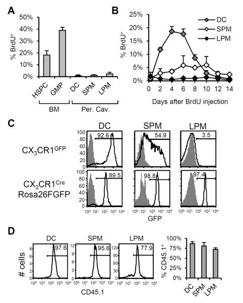
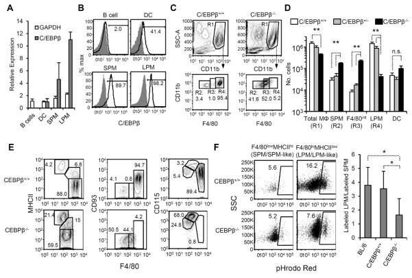
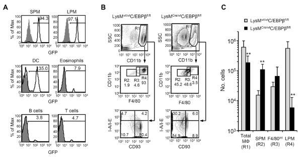
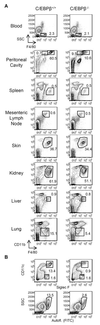
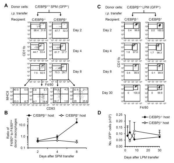
Macrophages and dendritic cells (DC) are distributed throughout the body and play important roles in pathogen detection and tissue homeostasis. In tissues, resident macrophages exhibit distinct phenotypes and activities, yet the transcriptional pathways that specify tissue-specific macrophages are largely unknown. We investigated the functions and origins of two peritoneal macrophage populations in mice: small and large peritoneal macrophages (SPM and LPM, respectively). SPM and LPM differ in their ability to phagocytose apoptotic cells, as well as in the production of cytokines in response to LPS. In steady-state conditions, SPM are sustained by circulating precursors, whereas LPM are maintained independently of hematopoiesis; however, both populations are replenished by bone marrow precursors following radiation injury. Transcription factor analysis revealed that SPM and LPM express abundant CCAAT/enhancer binding protein (C/EBP)-β. Cebpb(-/-) mice exhibit elevated numbers of SPM-like cells but lack functional LPM. Alveolar macrophages are also missing in Cebpb(-/-) mice, although macrophage populations in the spleen, kidney, skin, mesenteric lymph nodes, and liver are normal. Adoptive transfer of SPM into Cebpb(-/-) mice results in SPM differentiation into LPM, yet donor SPM do not generate LPM after transfer into C/EBPβ-sufficient mice, suggesting that endogenous LPM inhibit differentiation by SPM. We conclude that C/EBPβ plays an intrinsic, tissue-restricted role in the generation of resident macrophages.


Figures

Figure 1
Figure 1. Effector functions of SPM and LPM
(A) Flow cytometric gating strategy to identify peritoneal DC (R1: IgMCD11c+SSClow), SPM (R2: IgMCD11cCD11bhiF4/80low), and LPM (R3: IgMCD11cCD11bhiF4/80hi) in peritoneal lavages of C57BL/6 mice. Cells in gate R4 (IgMCD11cCD11bintSSChi) were identified as eosinophils. Forward- and side light scattering (FSC and SSC) properties and surface expression of CD11b, CD115, MHCII (I-A/I-E), CD93, and CD205 (open histograms) for each compartment is shown in histograms to the right. Gray histograms depict background staining by isotype-matched control antibodies. (B) Cytokine signatures of sorted peritoneal DC, SPM, and LPM following in vitro exposure to LPS. The concentrations of various cytokines in the supernatants were determined by multiplex bead array. Data are summarized here in a Venn diagram and detailed in Supplemental Figure 1. Data represent three independent experiments, n=3–7 samples per cell type. (C) Peritoneal cells were assessed for their ability to phagocytose apoptotic cells in vivo. pHrodo Red-labeled apoptotic thymocytes were injected i.p. and then peritoneal lavages were performed 1 hour later. Phagocytosis was measured as the frequency of pHrodo Red-labeled cells in the SPM, LPM, DC, and eosinophil compartments. The upper row shows representative dot plots of background fluorescence by cells of untreated mice, whereas the lower row depicts plots of gated cells from a mouse injected with labeled apoptotic cells. Data are representative of two individual experiments; n=4 mice.
Figure 2
Figure 2. Origins and maintenance of peritoneal DC, SPM, and LPM
(A) To measure proliferation in peritoneal myeloid compartments, C57BL/6 mice were injected i.p. with BrdU. Three hours later, peritoneal DC, SPM, and LPM were analyzed for BrdU uptake. Bone marrow hematopoietic stem and progenitor cells (HSPC: Linc-Kit+Sca-1+) and granulocyte/macrophage progenitors (GMP: Linc-Kit+Sca-1CD34+CD16/32+) were harvested as controls. The mean(±SD) frequency of BrdU+ cells in each compartment is shown. (B) BrdU transit through peritoneal DC, SPM, and LPM compartments after a single BrdU pulse. The mean(±SD) frequencies of BrdU+ cells in the DC (gray), SPM (white), and LPM (black) compartments at intervals after BrdU administration are shown. Data represent two independent experiments, n=2–4 mice per data point. (C) CX3CR1 expression by peritoneal DC, SPM, and LPM. In the top row, GFP fluorescence by cells from CX3CR1GFP/wt mice (open histogram) and C57BL/6 controls (shaded histogram) is shown. In the bottom row, GFP fluorescence by peritoneal DC, SPM, and LPM from CX3CR1Cre/wtRosa26R-FGFP mice (open histogram) and control CX3CR1wt/wtRosa26R-FGFP (shaded histogram) is shown. Histograms are representative of results in two independent experiments, n=3–4 mice per genotype. (D) Reconstitution of peritoneal DC, SPM, and LPM following irradiation and BM transfer. C57BL/6.CD45.1 BM cells were adoptively transferred into irradiated C57BL/6.CD45.2 hosts. Representative histograms show frequencies of donor-derived (CD45.1+) cells in peritoneal DC, SPM, and LPM compartments 4–5 weeks after reconstitution. At right, the mean(±SD) frequencies of CD45.1+ cells in each compartment are shown. Data represent two independent experiments, n=4 mice.
Figure 3
Figure 3. Effect of C/EBPβ deficiency on peritoneal macrophages
(A) Peritoneal B cells (IgM+), peritoneal DC, SPM, and LPM were sorted (gated as in Fig. 1A) and analyzed for C/EBPβ, GAPDH, and β-actin transcripts by quantitative RT-PCR. We measured C/EBPβ and GAPDH transcription relative to β-actin and then normalized to the average value of the peritoneal DC compartment. Values represent the geometric mean (±GSD) from three individual sorts. (B) C/EBPβ protein in SPM, LPM, peritoneal DC, and B cells. Intracellular staining of peritoneal cells with anti-C/EBPβ (open histogram) or isotype control (gray histogram) is shown. Data are representative of 2 individual experiments, n=2. (C) Representative flow cytometric analyses of peritoneal macrophages in C/EBPβ+/+ and C/EBPβ−/− mice. In the top row of dot plots, IgM+ and CD11c+ cells have been excluded as in Fig. 1A; SSChiCD11bhi cells are gated as total macrophages (R1: “Total MΦ”). F4/80 and CD11b expression by total macrophages of C/EBPβ+/+ and C/EBPβ−/− mice are shown in the bottom row, and populations have been subdivided by F4/80 staining intensity (R2: “SPM”, R3: “F4/80int”, and R4: “LPM”). (D) The mean(±SD) numbers of Total MΦ, F4/80low SPM, F4/80int macrophages, and F4/80hi LPM from C/EBPβ+/+(open bars), C/EBPβ+/− (gray bars), and C/EBPβ−/− (black bars) are shown. Data represent 9 independent experiments, n=6–8 mice per genotype. * P≤0.05, ** P≤0.01 vs. C/EBPβ+/+ mice. n.s., not significant. (E) Phenotypic analysis of total macrophages (R1) macrophages from C/EBPβ+/+ (top row) and C/EBPβ−/− (bottom row) mice. In all dot plots, F4/80 is shown on the x-axis and MHCII, CD93, and CD115 on the y-axes. Data are representative of 2–3 experiments, n≥3 mice per antibody panel. (F) The phagocytic capacity of C/EBPβ+/+ (top row) and C/EBPβ−/− (bottom row) macrophages was assessed following i.p. injection of pHrodo Red-labeled apoptotic thymocytes. Macrophages were gated based on F4/80 and MHCII expression as in the left panel of Fig. 3E. SPM and SPM-like macrophages were gated as F4/80lowMHCIIhi SPM; LPM and LPM-like macrophages were gated as F4/80hiMHCIIlow (C/EBPβ+/+ mice) and F4/80intMHCIIlow (C/EBPβ−/− mice) cells, respectively. The right panel shows the ratio (geometric mean±GSD) of labeled F4/80int/hiMHCIIlow macrophages to F4/80lowMHCIIhi macrophages in C57BL/6, C/EBPβ+/+ littermates, and C/EBPβ−/− mice. Flow plots are representative of 2 individual experiments, n=2–4 mice per genotype. * P≤0.05.
Figure 4
Figure 4. Abnormal distribution of peritoneal macrophage populations in LysMCre/WTC/EBPβfl/fl mice
(A) FGFP expression in SPM, LPM, DC, CD11bintSSChi eosinophils, IgM+ B cells, and TCRβ+ T cells in peritoneal lavages of LysMCre/WTRosa26R-FGFP mice (open histograms) and control LysMWT/WTRosa26R-FGFP mice (gray histograms). (B) Representative flow cytometric analyses of peritoneal macrophages in LysMwt/wtC/EBPβfl/fl and LysMCre/wtC/EBPβfl/fl mice. In the top row of dot plots, cells expressing IgM or CD11c have been excluded and SSChiCD11bhi cells gated as total macrophages (R1: “Total MΦ”). F4/80 and CD11b expression by total macrophages of C/EBPβ+/+ and C/EBPβ−/− mice is shown in the middle row, and populations have been subdivided by F4/80 staining intensity (R2: “SPM”, R3: “F4/80int”, and R4: “LPM”); CD93 and MHCII expression by total macrophages is shown in the lowest row. (C) The mean(±SD) numbers of Total MΦ, SPM, F4/80int macrophages, and LPM from LysMwt/wtC/EBPβfl/fl mice (gray bars) and LysMCre/wtC/EBPβfl/fl mice (black bars) are shown. Data represent two independent experiments, n=8–9 mice per genotype. ** P≤0.01.
Figure 5
Figure 5. Survey of tissue macrophages in C/EBPβ−/− mice
(A) Single-cell suspensions of blood, peritoneal cavity, spleen, mesenteric lymph nodes, skin (ear), kidney, liver, and lungs of C/EBPβ+/+ and C/EBPβ−/− mice were stained for CD45, F4/80, and CD11b. Viable CD45+ cells were gated for analysis of F4/80+ macrophage/monocyte populations. (B) Viable CD45+ lung cells from C/EBPβ+/+ and C/EBPβ−/− mice were analyzed for the presence of alveolar macrophages using Siglec F and CD11c (top row), and side-scatter (SSC) and autofluorescence (bottom row). Flow plots are representative of 2 individual experiments, n=3 mice per genotype.
Figure 6
Figure 6. SPM efficiently differentiate into LPM when adoptively transferred into C/EBPβ-deficient, but not -sufficient, hosts
(A) C/EBPβ+/+GFP+/+ SPM were isolated by cell sorting and injected i.p. into C/EBPβ-sufficient or -deficient hosts. Representative FACS plots of F4/80 and CD11b staining of GFP+IgMCD11c peritoneal cells recovered on days 2, 4, and 8 after transfer are shown. For day 8, MHCII and CD93 staining is shown for F4/80low and F4/80hi donor cells. (B) The ratio of F4/80hi cells to F4/80low cells was calculated in each mouse receiving GFP+ SPM. The geometric mean(±GSD) ratio of F4/80hi cells to F4/80low cells at each time point after transfer is shown. Data represent 4 independent experiments; n=2–4 mice per data point. (C) LPM from C/EBPβ+/+GFP+/+ mice were isolated by cell sorting and injected i.p. into C/EBPβ-sufficient or -deficient hosts. Representative FACS plots of F4/80 and CD11b staining of GFP+IgMCD11c cells on days 2, 4, 8, and 30 after transfer are shown. (D) The mean(±SD) numbers of GFP+ cells recovered in peritoneal lavages after adoptive transfer of C/EBPβ+/+GFP+/+ LPM into C/EBPβ-sufficient (C/EBPβ+/+ or C/EBPβ+/−) or -deficient hosts are shown. Data represent 5 independent experiments; n=2–4 mice per data point.

References

    1. van Furth R, Cohn ZA, Hirsch JG, Humphrey JH, Spector WG, Langevoort HL. The mononuclear phagocyte system: a new classification of macrophages, monocytes, and their precursor cells. Bull World Health Organ. 1972;46:845–852. - PMC - PubMed
    1. Gordon S, Taylor PR. Monocyte and macrophage heterogeneity. Nat Rev Immunol. 2005;5:953–964. - PubMed
    1. Geissmann F, Manz MG, Jung S, Sieweke MH, Merad M, Ley K. Development of monocytes, macrophages, and dendritic cells. Science. 2010;327:656–661. - PMC - PubMed
    1. Schulz C, Gomez Perdiguero E, Chorro L, Szabo-Rogers H, Cagnard N, Kierdorf K, Prinz M, Wu B, Jacobsen SE, Pollard JW, Frampton J, Liu KJ, Geissmann F. A lineage of myeloid cells independent of Myb and hematopoietic stem cells. Science. 2012;336:86–90. - PubMed
    1. Yona S, Kim KW, Wolf Y, Mildner A, Varol D, Breker M, Strauss-Ayali D, Viukov S, Guilliams M, Misharin A, Hume DA, Perlman H, Malissen B, Zelzer E, Jung S. Fate Mapping Reveals Origins and Dynamics of Monocytes and Tissue Macrophages under Homeostasis. Immunity. 2013;38:79–91. - PMC - PubMed

Publication types

MeSH terms

Substances

LinkOut - more resources