. 2013 Nov 1;191(9):4789-803.
doi: 10.4049/jimmunol.1300645. Epub 2013 Sep 27.

Proapoptotic chemotherapeutic drugs induce noncanonical processing and release of IL-1β via caspase-8 in dendritic cells

Affiliations

Proapoptotic chemotherapeutic drugs induce noncanonical processing and release of IL-1β via caspase-8 in dendritic cells

Christina Antonopoulos et al. J Immunol. .

Abstract

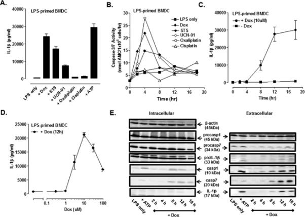
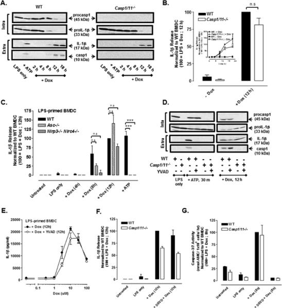
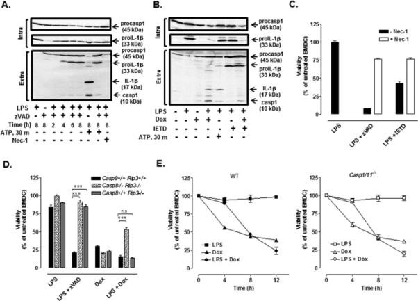
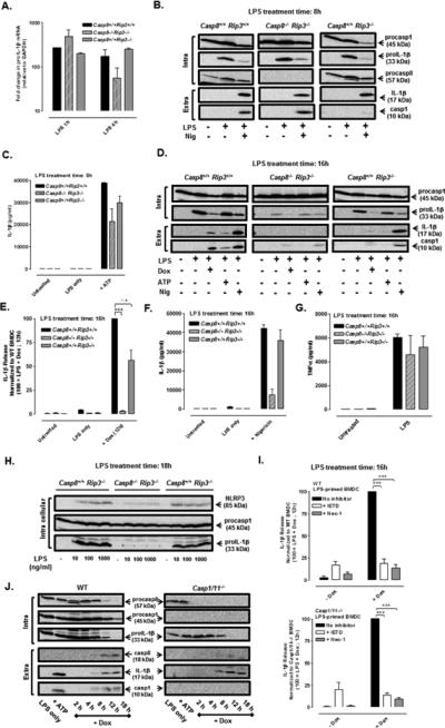
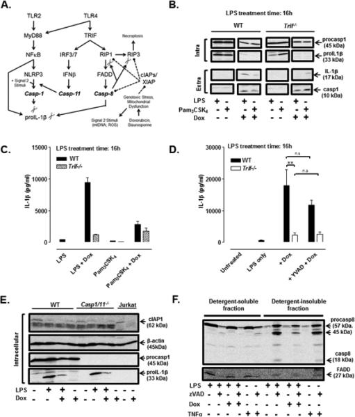
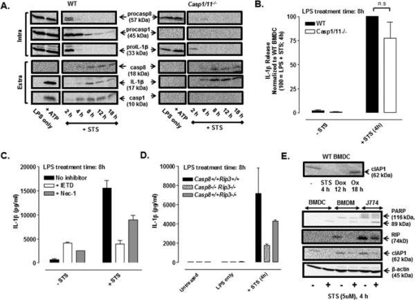
The identification of noncanonical (caspase-1-independent) pathways for IL-1β production has unveiled an intricate interplay between inflammatory and death-inducing signaling platforms. We found a heretofore unappreciated role for caspase-8 as a major pathway for IL-1β processing and release in murine bone marrow-derived dendritic cells (BMDC) costimulated with TLR4 agonists and proapoptotic chemotherapeutic agents such as doxorubicin (Dox) or staurosporine (STS). The ability of Dox to stimulate release of mature (17-kDa) IL-1β was nearly equivalent in wild-type (WT) BMDC, Casp1(-/-)Casp11(-/-) BMDC, WT BMDC treated with the caspase-1 inhibitor YVAD, and BMDC lacking the inflammasome regulators ASC, NLRP3, or NLRC4. Notably, Dox-induced production of mature IL-1β was temporally correlated with caspase-8 activation in WT cells and greatly suppressed in Casp8(-/-)Rip3(-/-) or Trif(-/-) BMDC, as well as in WT BMDC treated with the caspase-8 inhibitor, IETD. Similarly, STS stimulated robust IL-1β processing and release in Casp1(-/-)Casp11(-/-) BMDC that was IETD sensitive. These data suggest that TLR4 induces assembly of caspase-8-based signaling complexes that become licensed as IL-1β-converting enzymes in response to Dox and STS. The responses were temporally correlated with downregulation of cellular inhibitor of apoptosis protein 1, suggesting suppressive roles for this and likely other inhibitor of apoptosis proteins on the stability and/or proteolytic activity of the caspase-8 platforms. Thus, proapoptotic chemotherapeutic agents stimulate the caspase-8-mediated processing and release of IL-1β, implicating direct effects of such drugs on a noncanonical inflammatory cascade that may modulate immune responses in tumor microenvironments.


Figures

FIGURE 1
FIGURE 1. Pro-apoptotic chemotherapeutic drugs induce the release of IL-1β in LPS-primed murine bone marrow-derived dendritic cells (BMDC)
(A) BMDC were primed with LPS (1μg/ml) for 4 h prior to stimulation for 12 h with a panel of pro-apoptotic agents including staurosporine (STS, 5 μM), UCN-01 (10 μM), doxorubicin (Dox, 10 μM), oxaliplatin (Ox, 25 μM) and cisplatin (CDDP, 25 μM). The extracellular medium was collected and assayed for IL-1β by ELISA. BMDC were primed with LPS for 15.5 h prior to ATP (5mM) stimulation for 30 min. Results are the mean ± range of two experiments. (B) The kinetics of drug-induced caspase-3/7 activity in LPS-primed BMDC was measured by proteolytic cleavage of the DEVD-AMC substrate. The concentrations of each drug used were the same as described in (A). Results are from a single experiment. (C) The kinetics of IL-1β release from LPS-primed and Dox-stimulated (10 μM) WT BMDC were assayed by ELISA. Results are the mean ± SE from 4-8 experiments. (D) LPS-primed BMDC were stimulated with varying doses of Dox for 12 h. Results are the mean ± SE of 3 experiments. (E) WT BMDC were stimulated as in (C), and the extracellular medium and cell lysates were collected and processed for western blot analysis for detection of IL-1β, caspase-1, and caspase-7. BMDC were primed with LPS for 5.5 h prior to ATP (5mM) stimulation for 30 min. The data are representative of results from 3 experiments.
FIGURE 2
FIGURE 2. Doxorubicin induces caspase-1 independent processing and release of IL-1β in LPS-primed BMDC
(A) LPS-primed (1 μg/ml) WT or Casp1/11−/− BMDC were stimulated with Dox (10 μM) for 2-18 h, and western blot analysis of IL-1β and caspase-1 from cell lysates and extracellular supernatants was performed. BMDC were LPS-primed for 5.5 h followed by 30 min of ATP (5mM) stimulation. Results are representative of 3 identical experiments. (B) The release of IL-1β from LPS-primed (1ug/ml) WT or Casp1/11−/− BMDC stimulated or not with 10 μM Dox for 12 h was assayed by ELISA. IL-1β release was normalized to WT BMDC treated with LPS + Dox for 12 h and expressed as the mean ± SE of 5 experiments. The differences between WT and Casp1/11−/− BMDC were not significant (n.s, P > .05) by Student's t-test. Inset: Kinetics of Dox-induced IL-1β release (normalized to WT BMDC treated with LPS + Dox for 12 h) in LPS-primed WT versus Casp1/11−/− BMDC as described in part (A). Results are the mean ± SE of 3 experiments. (C) WT, Asc−/−, and Nlrp3−/− Nlrc4−/− BMDC were primed with LPS for 4 h before stimulation with Dox for 4, 8, or 12 h and assayed for release IL-1β release by ELISA. Parallel samples were primed with LPS for 15.5 h prior to ATP (5mM) stimulation for 30 min. IL-1β release was normalized to WT BMDC treated with LPS + Dox for 12 h and expressed as the mean ± SE of 3-5 experiments. ***P < .001 or n.s by ANOVA. (D) Western blot analysis of LPS-primed (1μg/ml) WT or Casp1/11−/− BMDC stimulated with 10 μM Dox for 12 h or 5mM ATP for 30 min in the presence or absence of the caspase-1 inhibitor, YVAD (50 μM). BMDC were LPS-primed for 7.5 h followed by ATP (5mM) stimulation for 30 min. The data are representative of results from 2 experiments. (E) Concentration-response relationship for Dox-stimulated IL-1β release in the presence or absence of YVAD in LPS-primed BMDC. The curve depicted without YVAD treatment is the same as shown in Figure 1D. Results are the mean ± SE of 3 experiments. (F) The release of IL-1β was measured in LPS-primed WT or Casp1/11−/− BMDC stimulated ± 10μM Dox for 12 h in the presence or absence of DEVD (50μM). IL-1β release was normalized to WT BMDC treated with LPS + Dox for 12 h. Results are the mean ± range of 2 experiments. (G) Caspase-3/7 activity was measured in LPS-primed WT and Casp1/11−/− BMDC ± 10 μM Dox in the presence or absence of DEVD. Casp3/7 activity was normalized to WT BMDC treated with LPS + Dox for 8 h. Results are the mean ± range of 2 experiments.
FIGURE 3
FIGURE 3. TLR4 activation coupled with caspase-8 inhibition induces RIP1/RIP3-dependent necroptosis and release of unprocessed proIL-1β while TLR4 activation coupled with Dox treatment induces caspase-8-dependent apoptosis
(A) Western blot analysis of IL-1β and caspase-1 in cell lysates and extracellular medium from WT BMDC treated with LPS (1μg/ml) and zVAD (50 μM) for 2-8 h with or without necrostatin-1 (Nec-1, 50 μM). (B) Western blot analysis of IL-1β and caspase-1 in cell lysates and extracellular medium from WT BMDC treated with LPS for 4 h prior to stimulation with Dox (10 μM) in the presence or absence of IETD (100uM) for 12 h. In panels A and B, BMDC were primed with LPS for 7.5 h prior to 30 min of ATP (5mM) stimulation. The data are representative of results from 2-3 experiments. (C) WT BMDC were co-treated with LPS + zVAD or LPS + IETD in the presence or absence of Nec-1, and cell viability was assessed by measuring intracellular ATP content. Results are from a single experiment with each condition performed in quadruplicate. (D) WT, Casp8−/− Rip3−/−, and Casp8+/+ Rip3−/− BMDC were treated with LPS (16 h), Dox (12h), or LPS for 4 h prior to Dox or zVAD for 12 h, and cell viability was assessed. Results are from a representative experiment (of 2 similar experiments) performed in quadruplicate. ***P < .001 by ANOVA. (E) WT and Casp1/11−/− BMDC were LPS-primed or not for 4 h prior to stimulation or not with Dox for 4, 8, or 12 h, and cell viability was assessed. Results are from a representative experiment of 2 similar experiments with each condition performed in triplicate.
FIGURE 4
FIGURE 4. Doxorubicin induces caspase-8 dependent IL-1β processing and release in LPS-primed BMDC
(A) ProIL-1β mRNA (normalized to GAPDH) was measured by qPCR in WT, Casp8−/− Rip3−/−, and Casp8+/+ Rip3−/− BMDC treated with LPS (1 μg/ml) for 1 h or 4 h. Results are from a single experiment. (B) Western blot analysis of proIL-1β, procaspase-1, and procaspase-8 in cell lysates (intra) and mature IL-1β and caspase-1 p10 subunit in extracellular (extra) supernatants of WT, Casp8−/− Rip3−/−, and Casp8+/+ Rip3−/− BMDC treated with LPS for 8 h and stimulated ± nigericin (10 μM) for the final 30 min of the LPS treatment period. Data are representative of results from 2 experiments. (C) BMDC of the indicated genotypes were treated with LPS (1 μg/ml) for 7.5 h prior to simulation ± ATP (5 mM) for an additional 30 min; IL-1β release was assayed by ELISA. Results are the mean ± range of 2 experiments. (D) Western blot analysis of pro-IL-1β and procaspase-1 in cell lysates (intra) and mature IL-1β and caspase-1 p10 subunit in extracellular (extra) supernatants of WT, Casp8−/− Rip3−/−, and Casp8+/+ Rip3−/− BMDC treated with LPS for a total of 16 h and stimulated with either Dox (10 μM) for the final12 h, or with ATP (5 mM) or nigericin (10 μM) for the final 30 min, of the LPS treatment period . Results are representative of 3 similar experiments. (E, F) WT, Casp8−/− Rip3−/−, and Casp8+/+ Rip3−/− BMDC were treated with LPS (1 μg/ml) for a total of 16 h and stimulated with Dox (10 μM) for the last 12 h (E) or nigericin (10 μM) for the last 30 min (F) of the LPS treatment period. IL-1β release was assayed by ELISA and normalized to WT stimulated with LPS + Dox (E) or WT stimulated with LPS + nigericin (F). Results are the mean ± SE of 3 experiments for (E) or the mean ± SE for 2 experiments (F) ***P < .001 or not significant (n.s) by ANOVA. (G) TNFα release (by ELISA) from WT, Casp8−/− Rip3−/−, and Casp8+/+ Rip3−/− BMDC treated ± LPS (1 μg/ml) for 16 h. Results are the mean ± range of 2 experiments. (H) Western blot analysis of proIL-1β, procaspase-1, and NLRP3 expression in WT, Casp8−/− Rip3−/−, and Casp8+/+ Rip3−/− BMDC treated for 18 h with 0, 10, 100, or 1000 ng/ml of LPS. Results are representative of 2 experiments. (I) WT and Casp1/11−/− BMDC were treated with LPS (1 μg/ ml) for a total of 16 h and stimulated ± Dox (10 μM), ± IETD (100 μM), ± Nec-1 (50 μM) for the final 12 h of the LPS treatment period. IL-1β release was assayed by ELISA and normalized to the samples stimulated with LPS + Dox. Results are the mean ± SE of 3 experiments. ***P < .001 by ANOVA. (J) Western blot analysis of procapase-8, procaspase-1, and proIL-1β in cell lysates (intra) and mature IL-1β, caspase-8 p18 subunit, and caspase-1 p10 subunit in the extracellular supernatants (extra) from WT or Casp1/11−/− BMDC treated with LPS (1 μg/ml) for 4 h prior to co-stimulation with Dox (10 μM) for another 2-18 h or with LPS for 5.5 h prior to ATP (5mM) stimulation for 30 min. The data are representative of results from 3 experiments.
FIGURE 5
FIGURE 5. LPS + Dox-induced IL-1β release is TRIF-dependent and correlated with IAP downregulation and recruitment of caspase-8/ FADD to a detergent-insoluble compartment
(A) A model for parallel pathways of canonical NLRP3 inflammasome activation and TRIF-induced caspase-8 signaling complexes that may mediate IL-1β processing induced by LPS + doxorubicin. (B) Western blot analysis of proIL-1β and procaspase-1 in cell lysates (intra) and mature IL-1β and caspase-1 p10 subunit in extracellular supernatants (extra) from WT and Trif−/− BMDC stimulated Pam3CSK4 (200 ng/ml) ± Dox (10 μM) or LPS (1 μg/ml) ± Dox for 12 h. The data are representative of 2 experiments. (C, D) IL-1β release (by ELISA) from WT or Trif−/− BMDC treated with Pam3CSK4 (200 ng/ml) or LPS (1 μg/ml) for a total of 16 h and stimulated ± Dox (10 μM) (C, D) or Dox + YVAD (50 μM) (D) for the final 12 h of the LPS or Pam3CSK4 treatment periods). Data in (C) are representative of results from 2 experiments. Data in (D) are the mean ± SE of 3 experiments. **P < .01 or not significant (n.s) by ANOVA. (E) Western blot analysis of cIAP1, proIL-1β, procaspase-1, and actin in cell lysates from WT BMDC or Casp1/11−/− BMDC treated ± LPS (1 μg/ml) for 4 h prior to stimulation ± Dox (10 μM) for 12 h. As a positive control, lysates from control or Dox-treated Jurkat leukemic T cells were also analysed. Results are from a single experiment. (F) WT BMDC were treated with LPS (1 μg/ml) or TNFα (50 ng/ml) for 2 h in the presence or absence of zVAD (50 μM). Alternatively, BMDC were treated with LPS for 4 h prior to stimulation with Dox (10 μM) or Dox + zVAD for another 8 h. Cell lysates were separated into detergent-soluble versus detergent-insoluble fractions for western blot analysis of caspase-8 and FADD. Results are representative of 2 experiments.
FIGURE 6
FIGURE 6. Staurosporine induces caspase-8-dependent IL-1β processing and release in LPS-primed BMDC
(A) Western blot analysis of procapase-8, procaspase-1, and proIL-1β in cell lysates (intra) and mature IL-1β, caspase-8 p18 subunit, and caspase-1 p10 subunit in the extracellular supernatants (extra) from WT or Casp1/11−/− BMDC were treated ± LPS (1 μg/ml) for 4 h and then co-stimulated with STS (5 μM) for an additional 2-18 h or with LPS for 5.5 h prior to ATP (5mM) stimulation for 30 min. The data are representative of results from 3 experiments. (B) LPS-primed (1 μg/ml, 4h) WT and Casp1/11−/− BMDC were stimulated ± STS (5 μM) for 4 h; IL-1β release was assayed by ELISA and normalized to WT BMDC treated with LPS + STS (4 h). Results are the mean ± SE of 4-5 experiments. The differences between WT and Casp1/11−/− BMDC were not significant (n.s, P > .05) by Student's t-test. (C) WT BMDC were primed with LPS (1 μg/ml, 4 h) prior to stimulation ± STS (5 μM, 4 h) in the presence or absence of IETD (100 μM) or Nec-1 (50 μM); IL-1β release was assayed by ELISA. Results are from a single representative experiment of 3 experiments. (D) WT, Casp8−/− Rip3−/−, and Casp8+/+ Rip3−/− BMDC were treated ± LPS (1 μg/ml) for 4 h prior to co-stimulation ± STS (5 μM) for an additional 4 h. Results are from a single experiment with each condition was performed in duplicate. (E) Upper panel: Western blot analysis of cIAP1 of lysates from untreated WT BMDC or WT BMDC treated with STS (5 μM, 4 h), Dox (10 μM, 12 h), or oxaliplatin (Ox) (25 μM, 18 h). Lower panel: Western blot analysis of cIAP1, PARP (full length and cleaved forms), and RIP1 in lysates in control versus STS-treated (5 μM,4 h) BMDC, BMDM, and J774 murine macrophages. Results are from a single experiment.

References

    1. Casares N, Pequignot MO, Tesniere A, Ghiringhelli F, Roux S, Chaput N, Schmitt E, Hamai A, Hervas-Stubbs S, Obeid M, Coutant F, Metivier D, Pichard E, Aucouturier P, Pierron G, Garrido C, Zitvogel L, Kroemer G. Caspase-dependent immunogenicity of doxorubicin-induced tumor cell death. J. Exp. Med. 2005;202:1691–1701. - PMC - PubMed
    1. Tesniere A, Schlemmer F, Boige V, Kepp O, Martins I, Ghiringhelli F, Aymeric L, Michaud M, Apetoh L, Barault L, Mendiboure J, Pignon JP, Jooste V, van Endert P, Ducreux M, Zitvogel L, Piard F, Kroemer G. Immunogenic death of colon cancer cells treated with oxaliplatin. Oncogene. 2010;29:482–491. - PubMed
    1. Rathinam VA, Vanaja SK, Fitzgerald KA. Regulation of inflammasome signaling. Nat. Immunol. 2012;13:333–332. - PMC - PubMed
    1. Shimada K, Crother TR, Karlin J, Dagvadorj J, Chiba N, Chen S, Ramanujan VK, Wolf AJ, Vergnes L, Ojcius DM, Rentsendorj A, Vargas M, Guerrero C, Wang Y, Fitzgerald KA, Underhill DM, Town T, Arditi M. Oxidized mitochondrial DNA activates the NLRP3 inflammasome during apoptosis. Immunity. 2012;36:401–414. - PMC - PubMed
    1. Perregaux D, Gabel CA. Interleukin-1 beta maturation and release in response to ATP and nigericin. Evidence that potassium depletion mediated by these agents is a necessary and common feature of their activity. J. Biol. Chem. 1994;269:15195–15203. - PubMed

Publication types

MeSH terms

Substances