

Clinical Cerenkov luminescence imaging of (18)F-FDG
- PMID: 24078721
- PMCID: PMC3903390
- DOI: 10.2967/jnumed.113.127266
Clinical Cerenkov luminescence imaging of (18)F-FDG
Abstract
The aim of this study was to determine the feasibility of Cerenkov luminescence (CL) imaging of patients undergoing diagnostic (18)F-FDG scans to detect nodal disease.
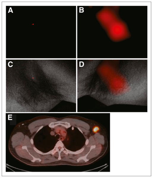
Methods: Patients undergoing routine (18)F-FDG PET/CT for various malignancies consented to being scanned for CL. White-light and Cerenkov images (5-min acquisition) of the surface of the patient contralateral to and at the site of nodal (18)F-FDG uptake were acquired using a cooled, intensified charge-coupled-device camera.
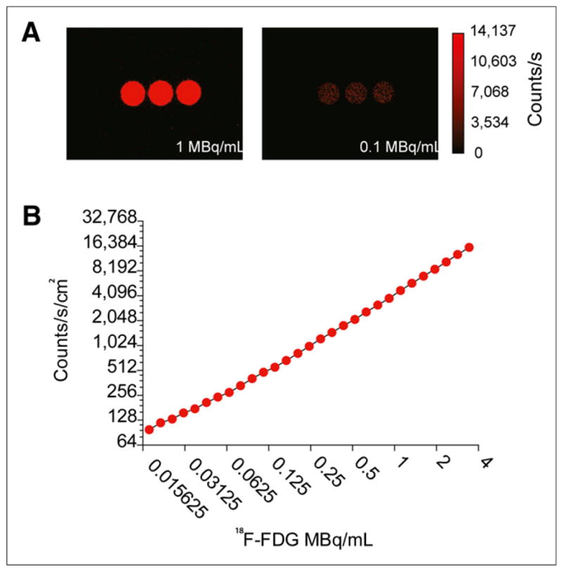
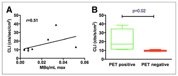
Results: The camera demonstrated linear correlation between activity and counts into the low nanocurie range using (18)F-FDG. Imaging of patients revealed the presence of (18)F-FDG uptake in nodes that demonstrated uptake on PET. A correlation between maximum standardized uptake value from PET and counting rate per area on the CL imaging was established.
Conclusion: CL imaging with diagnostic doses of (18)F-FDG is feasible and can aid in detecting disease in the clinical setting.
Keywords: 18F-FDG; Cerenkov luminescence imaging; PET/CT; clinical.
Conflict of interest statement
No other potential conflict of interest relevant to this article was reported.
Figures
Comment in
-
Reply: Human Cerenkov imaging using 18F-FDG.J Nucl Med. 2014 Mar;55(3):523-4. doi: 10.2967/jnumed.113.135533. Epub 2014 Jan 21. J Nucl Med. 2014. PMID: 24449595 No abstract available.
-
Human Cerenkov imaging using 18F-FDG.J Nucl Med. 2014 Mar;55(3):523. doi: 10.2967/jnumed.113.135384. Epub 2014 Jan 21. J Nucl Med. 2014. PMID: 24449596 No abstract available.
References
-
- Ntziachristos V, Ripoll J, Wang LV, Weissleder R. Looking and listening to light. Nat Biotechnol. 2005;23:313–320. - PubMed
-
- Willmann JK, van Bruggen N, Dinkelborg LM, Gahbhir SS. Molecular imaging in drug development. Nat Rev Drug Discov. 2008;7:591–607. - PubMed
-
- Byrne WL, Delille A, Kuo C, et al. Use of optical imaging to progress novel therapeutics to the clinic. J Control Release. 2013 May 13; Epub ahead of print. - PubMed
-
- Spinelli AE, D’Ambrosio D, Calderan L, Marengo M, Sbarbati A, Boschi F. Cerenkov radiation allows in vivo optical imaging of positron emitting radio-tracers. Phys Med Biol. 2010;55:483–495. - PubMed
Publication types
MeSH terms
Substances
Grants and funding
LinkOut - more resources
Full Text Sources
Other Literature Sources
Medical