. 2013:2013:650812.
doi: 10.1155/2013/650812. Epub 2013 Sep 2.

SOCS3 expression correlates with severity of inflammation, expression of proinflammatory cytokines, and activation of STAT3 and p38 MAPK in LPS-induced inflammation in vivo

Affiliations

SOCS3 expression correlates with severity of inflammation, expression of proinflammatory cytokines, and activation of STAT3 and p38 MAPK in LPS-induced inflammation in vivo

João Antônio Chaves de Souza et al. Mediators Inflamm. 2013.

Abstract

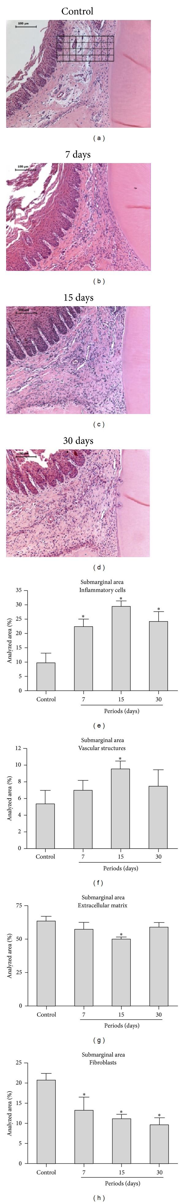
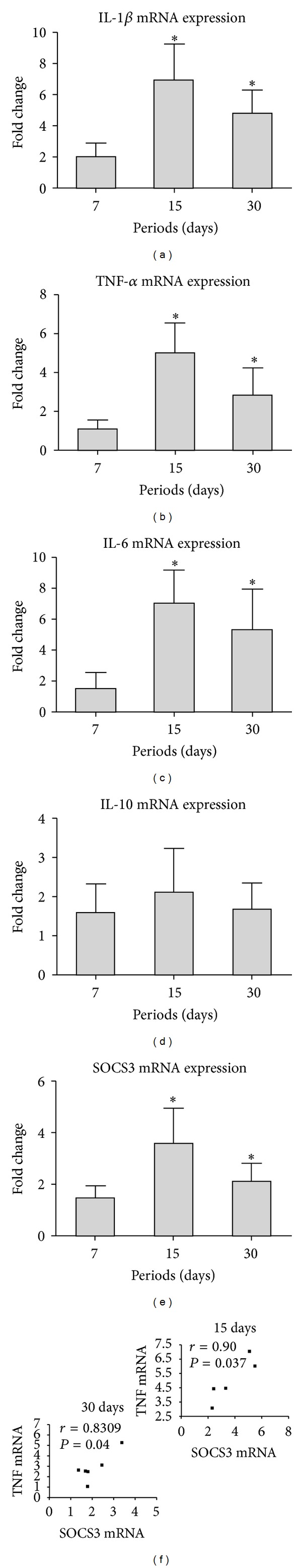
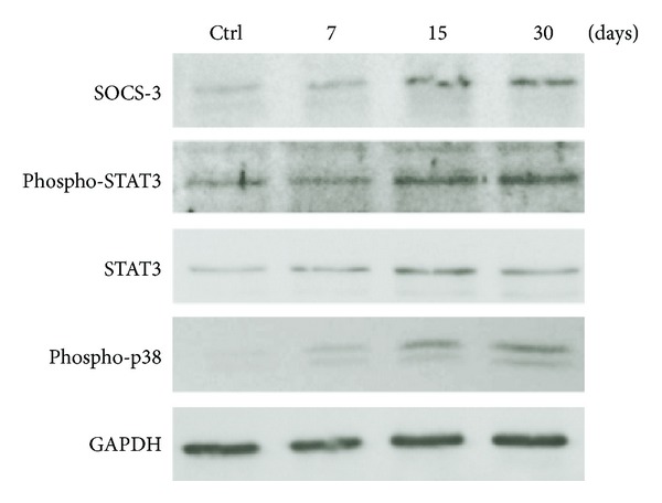
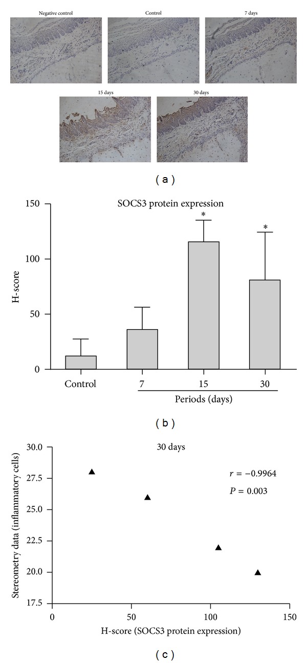
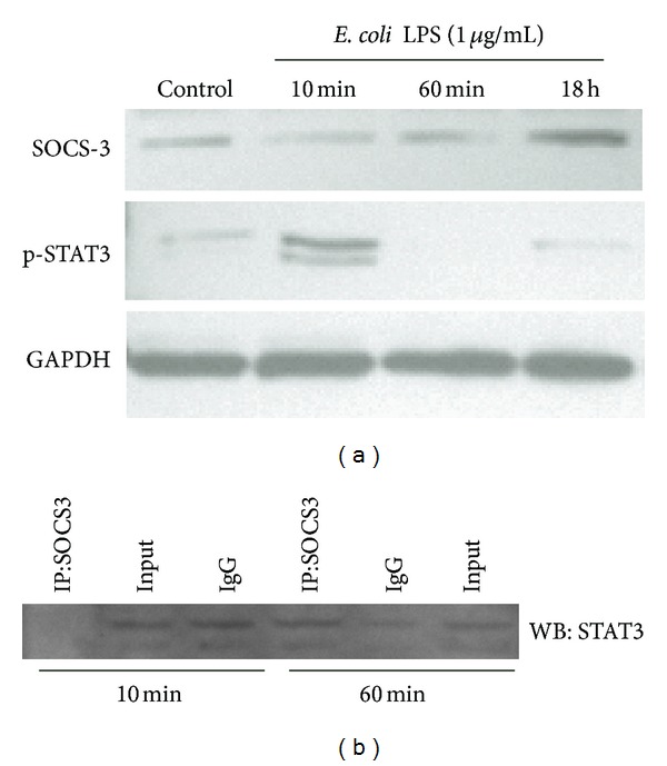
SOCS3 is an inducible endogenous negative regulator of JAK/STAT pathway, which is relevant in inflammatory conditions. We used a model of LPS-induced periodontal disease in rats to correlate SOCS3 expression with the inflammatory status. In vitro we used a murine macrophage cell line to assess the physical interaction between SOCS3 and STAT3 by coimmunoprecipitation. 30 ug of LPS from Escherichia coli were injected in the gingival tissues on the palatal aspect of first molars of the animals 3x/week for up to 4 weeks. Control animals were injected with the vehicle (PBS). The rats were sacrificed at 7, 15, and 30 days. Inflammation and gene expression were assessed by stereometric analysis, immunohistochemistry, RT-qPCR, and western blot. LPS injections increased inflammation, paralleled by an upregulation of SOCS3, of the proinflammatory cytokines IL-1 β , IL-6, and TNF- α and increased phosphorylation of STAT3 and p38 MAPK. SOCS3 expression accompanied the severity of inflammation and the expression of proinflammatory cytokines, as well as the activation status of STAT3 and p38 MAPK. LPS stimulation in a macrophage cell line in vitro induced transient STAT3 activation, which was inversely correlated with a dynamic physical interaction with SOCS3, suggesting that this may be a mechanism for SOCS3 regulatory function.


Figures

Figure 1
Figure 1
Representative images and stereometric analysis of each experimental period (7, 15, and 30 days) for the LPS-injected tissues (experimental group). A single representative image of the control group is shown, as no important changes were noted in this group in the different experimental periods (data not shown). The general characteristics of the inflammatory reaction include increase in inflammatory-like cell density and vascular structures, and a decrease in the number of fibroblast-like cells and extracellular matrix content was observed in all experimental periods. The image of a control site depicts the placement of the 3240 μm2 grids on the submarginal area limited coronally by the apical border of the junctional epithelium and laterally by the tooth structure. Stereometric analysis indicates that LPS injections caused a sustained inflammatory reaction. The relative presence of inflammatory cells, vascular structures, fibroblasts, and extracellular matrix in 32400 μm2 area (schematically shown on (a)) was analyzed. The severity of inflammation was significantly higher in comparison to the control group throughout the 30-day experimental period. *Significant differences compared with control groups (P < 0.05, ANOVA, and post hoc Tukey for pairwise comparisons).
Figure 2
Figure 2
Profile of the gene expression of SOCS3, proinflammatory cytokines IL-1β, TNF-α, and IL-6 and anti-inflammatory cytokine IL-10 during the course of LPS-induced periodontal disease. Expression of all proinflammatory cytokines and SOCS3 was increased at the 15- and 30-day experimental periods, with a peak in this increase at 15 days. No regulation of the anti-inflammatory cytokine IL-10 was observed in any of the experimental periods. mRNA expression was normalized to the expression of the housekeeping gene GAPDH. The bars represent mean fold changes, and the vertical lines represent the standard deviation of the mean fold change of six animals in each experimental group in comparison to untreated control. *Significant differences (P < 0.05, ANOVA, and post hoc Tukey tests for pairwise comparisons).
Figure 3
Figure 3
Western blot analysis of SOCS3, STAT3, and p38 MAPK protein expression in the LPS model of periodontal disease. Total protein was extracted from gingival tissue samples obtained from LPS and control sites at 7-, 15-, and 30-day periods. Activation of STAT3 and p38 as well as the expression of SOCS3 was determined. Increases in expression of SOCS3 and in the activation of STAT3 and p38 MAPK were observed at 7-, 15-, and 30-day experimental periods. Note that total STAT3 (including phosphorylated and nonphosphorylated forms) was also increased in the experimental group. The images are representative of the results obtained using samples from three different animals per period.
Figure 4
Figure 4
Immunohistochemical analysis revealed an increased number of SOCS3-positive cells after 7, 15, and 30 days after LPS injection. This increase reached statistical significance in comparison with gingival tissues of the control group at 15 and 30 days, as indicated by H-score analysis. Note that SOCS3-positive cells were located primarily near blood vessels and on the surface of alveolar bone. *Significant differences compared with control groups (P < 0.05).
Figure 5
Figure 5
LPS stimulation transiently activates STAT3 in raw 264.7 macrophages. Expression of SOCS3 was increased only after 18 h of LPS stimulation (a). Coimmunoprecipitation indicates a dynamic direct physical interaction between SOCS3 and STAT3 in murine macrophages. Raw 264.7 cells were stimulated with LPS (or the same volume of PBS vehicle) for 10 and 60 min. SOCS3 was immunoprecipitated from 500 ug of cell lysates, and after electrophoretic separation (SDS-PAGE), STAT3 was detected by western blotting. “Input” indicates the same quantity of cell lysate loaded onto “empty” affinity columns (no primary antibody immobilized), and “IgG” represents columns in which the same quantity of irrelevant IgG from rabbit was immobilized in the columns. Image is representative of three independent experiments.

References

    1. Taubman MA, Valverde P, Han X, Kawai T. Immune response: they key to bone resorption in periodontal disease. Journal of Periodontology. 2005;76(supplement 11):2033–2041. - PubMed
    1. Souza PPC, Palmqvist P, Lundgren I, et al. Stimulation of IL-6 cytokines in fibroblasts by toll-like receptors 2. Journal of Dental Research. 2010;89(8):802–807. - PubMed
    1. Liu YG, Lerner UH, Teng YA. Cytokine responses against periodontal infection: protective and destructive roles. Periodontology 2000. 2010;52(1):163–206. - PubMed
    1. Starr R, Hilton DJ. Negative regulation of the JAK/STAT pathway. Bioessays. 1999;21(1):47–52. - PubMed
    1. Alexander WS, Hilton DJ. The role of Suppressors of Cytokine Signaling (SOCS) proteins in regulation of the immune response. Annual Review of Immunology. 2004;22:503–529. - PubMed

Publication types

Substances

LinkOut - more resources