

Degenerative lumbar spinal stenosis in older people: current treatment options
- PMID: 24078855
- PMCID: PMC3784039
- DOI: 10.3238/arztebl.2013.0613
Degenerative lumbar spinal stenosis in older people: current treatment options
Abstract
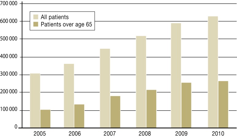
Background: Degenerative lumbar spinal stenosis is increasingly being diagnosed in persons over age 65. In 2011, 55 793 older people with this condition were treated as inpatients in German hospitals. Among physicians, there is much uncertainty about the appropriate treatment strategy.
Method: Selective literature review.
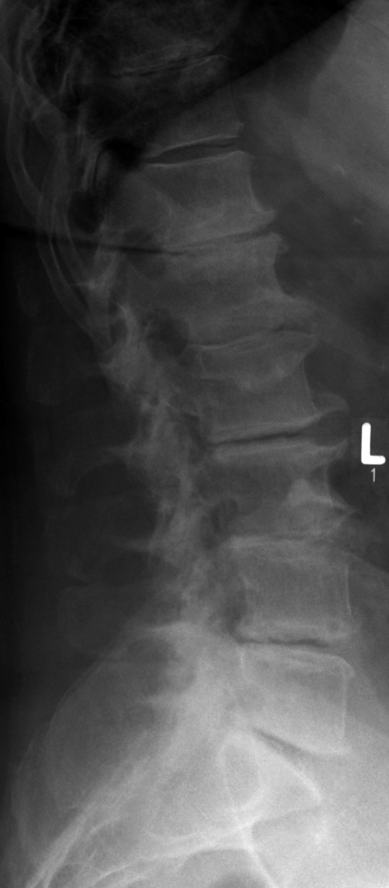
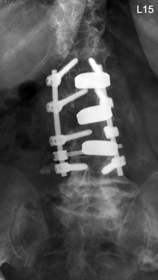
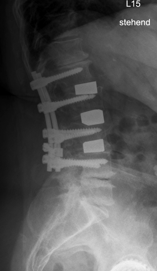
Results: Lumbar spinal stenosis in older people is characterized by spinal claudication and neurological deficits. A precise clinical history and physical examination and ancillary radiological studies are the necessary prerequisites for treatment. Magnetic resonance imaging is the radiological study of choice. Conservative treatment consists of physiotherapy, drugs, and local injections; various surgical treatments can be considered, depending on the severity of the problem. The main purpose of surgery is to decompress the spinal canal. If the lumbar spine is demonstrably unstable, an instrumented fusion should be performed in addition. There is, however, only moderately good evidence supporting the superiority of surgery over conservative treatment. In a prospective study, the complication rate of purely decompressive surgery was found to be 18%. The utility of the current operative techniques cannot be definitively assessed, because they are applied to a wide variety of patients in different stages of the disease and at different degrees of severity, and the reported results are thus not comparable from one trial to another.
Conclusion: No evidence-based recommendation on the diagnosis and treatment of lumbar spinal stenosis in older people can be formulated at present because of the lack of pertinent randomized trials.
Figures
Comment in
-
Further aspects of the therapeutic options.Dtsch Arztebl Int. 2014 Jan 17;111(3):39. doi: 10.3238/arztebl.2014.0039a. Dtsch Arztebl Int. 2014. PMID: 24606787 Free PMC article. No abstract available.
-
Methods need to be adapted to problems.Dtsch Arztebl Int. 2014 Jan 17;111(3):39. doi: 10.3238/arztebl.2014.0039b. Dtsch Arztebl Int. 2014. PMID: 24606788 Free PMC article. No abstract available.
-
In reply.Dtsch Arztebl Int. 2014 Jan 17;111(3):40. doi: 10.3238/arztebl.2014.0040. Dtsch Arztebl Int. 2014. PMID: 24606789 Free PMC article. No abstract available.
References
-
- Allen RT, Rihn JA, Glassman SD, Currier B, Albert TJ, Phillips FM. An evidence-based approach to spine surgery. Am J Med Qual. 2009;24(Suppl 6):15–24. - PubMed
-
- Watters WC, Baisden J, Gilbert TJ, et al. Degenerative lumbar spinal stenosis: an evidence-based clinical guideline for the diagnosis and treatment of degenerative lumbar spinal stenosis. Spine J. (3rd) 2008;8:305–310. - PubMed
-
- Statistisches Bundesamt Gesundheit. Fachserie 12 Reihe 6.2.1. Wiesbaden: Statistisches Bundesamt; 2011. Diagnosedaten der Patienten und Patientinnen in Krankenhäusern (einschließlich Sterbe- und Stundenfälle)
-
- Cheung KM, Karppinen J, Chan D, et al. Prevalence and pattern of lumbar magnetic resonance imaging changes in a population study of one thousand forty-three individuals. Spine. 2009;34:934–940. - PubMed
-
- Jensen MC, Brant-Zawadzki MN, Obuchowski N, Modic MT, Malkasian D, Ross JS. Magnetic resonance imaging of the lumbar spine in people without back pain. N Engl J Med. 1994;331:69–73. - PubMed
Publication types
MeSH terms
LinkOut - more resources
Full Text Sources
Other Literature Sources
Medical