. 2012;1(2):95-106.
doi: 10.4161/intv.22823.

In vivo dynamics of innate immune sentinels in the CNS

Affiliations

In vivo dynamics of innate immune sentinels in the CNS

Debasis Nayak et al. Intravital. 2012.

Abstract

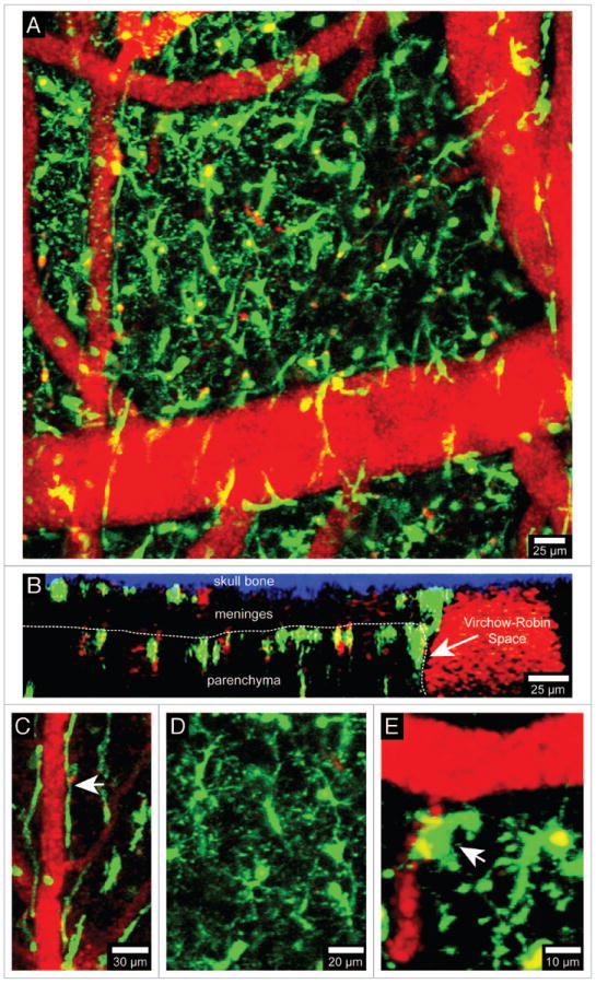
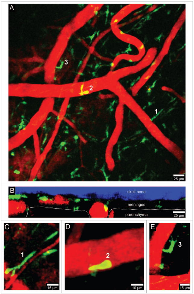
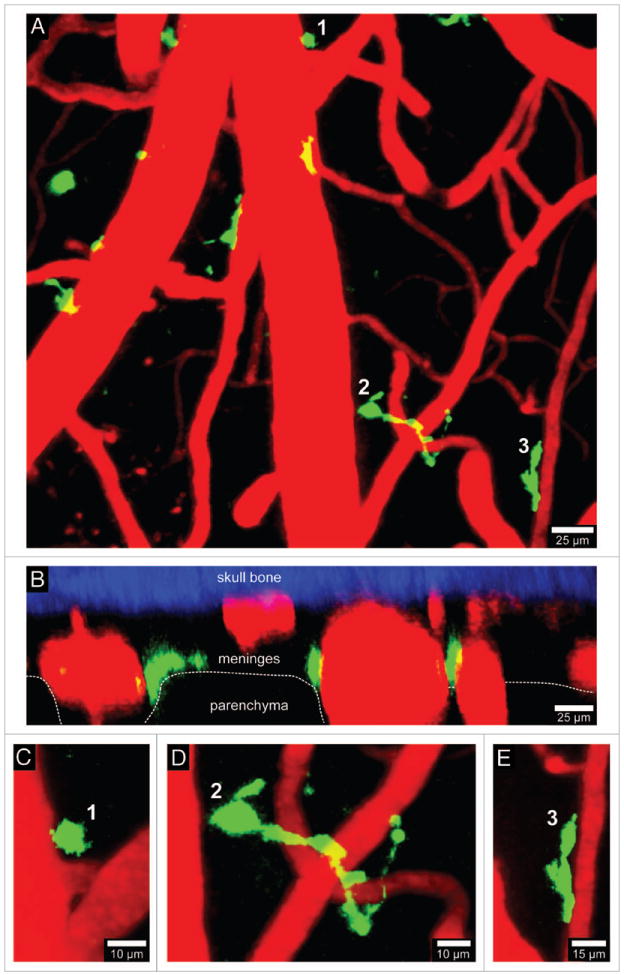
The innate immune system is comprised of cellular sentinels that often serve as the first responders to injury and invading pathogens. Our basic understanding of innate immunity is derived from research conducted in peripheral lymphoid tissues. However, it is now recognized that most non-lymphoid tissues throughout the body are equipped with specialized innate immune cells that are uniquely adapted to the niches in which they reside. The central nervous system (CNS) is a particularly interesting compartment because it contains a population of post-mitotic cells (neurons) that are intolerant of robust, cytopathic inflammatory responses observed in many peripheral tissues. Thus, evolutionary adaptations have fitted the CNS with a unique array of innate immune sentinels that facilitate the development of local inflammatory responses but attempt to do so in a manner that preserves the integrity of its post-mitotic residents. Interestingly, studies have even suggested that CNS resident innate immune cells contribute to the homeostasis of this compartment and promote neural activity. In this review we discuss recent advances in our understanding of CNS innate immune sentinels and how novel imaging approaches such as intravital two-photon laser scanning microscopy (TPLSM) have shed light on these cells during states of health and disease.

Keywords: CNS; dendritic cells; innate immunity; macrophages; microglia.


Conflict of interest statement

Disclosure of Potential Conflicts of Interest

No potential conflicts of interest were disclosed.

Figures

Figure 1
Figure 1
Visualization of brain myeloid cells in CX3CR1-GFP mice. TPLSM was used to capture 4D time-lapses through a thinned skull window of a naïve CX3CR1-GFP+/- mouse. The bone was thinned down manually to a thickness of ~30 μm and then imaged using a Leica SP5 two-photon microscope fitted with a 20x water dipping objective (1.0 NA). For imaging, the lens was dipped into artificial cerebral spinal fluid placed on top of the thinned skull window. Images were collected with a 1.0 μm step size to a depth of 100 μm beneath the skull surface. Z stacks were acquired every minute. Panel A shows the xy distribution of innate myeloid cells (green) in the meninges and neocortex of a naïve mouse brain. Blood vessels (red) were labeled by injecting 655-nm quantum dots intravenously before imaging. Panel B shows an xz projection from the same image stack in which the skull bone (blue), meninges, and brain parenchyma are visible. The white dotted line denotes the glial limitans. The Virchow-Robin space adjacent to a large blood vessel is also visible (white arrow). Note that the density of innate myeloid cells is greater in the parenchyma than in the meninges. CX3CR1-GFP+/- mice can be used to visualize meningeal macrophages (C, white arrow), microglia (D), and perivascular macrophages (E, white arrow). Meningeal macrophages (C) are worm-like cells that line blood vessels in the meninges. Perivascular macrophages (E) reside in Virchow-Robin spaces and adjacent to blood vessels found the brain parenchyma (white arrow). Microglia (D) are the most common CNS myeloid cell and are distributed uniformly throughout the brain parenchyma. Note that microglia are highly ramified cells, whereas meningeal and perivascular macrophages are not. See Video S1.
Figure 2
Figure 2
Visualization of peripherally-derived myelomonocytic cells in LysM-GFP mice. A 3D time-lapse was captured through a thinned skull window of a naïve LysM-GFP mouse in a manner similar to that described in Figure 1. Monocytes, macrophages, and neutrophils (but not microglia) are visible in this transgenic mouse strain. Panel A (xy maximal projection) shows the distribution of myelomonocytic cells (green) in relation to blood vessels (red). Note in B (xz projection) that the LysM-GFP+ cells reside exclusively in the meningeal and perivascular spaces. Three cellular morphologies are depicted in panels C–E (labeled 1–3). Worm-like meningeal macrophages (C) are visible along meningeal blood vessels similar to those seen in CX3CR1-GFP+/- mice. On occasion, neutrophils / monocytes (D) can be observed patrolling blood vessels. In addition, amoeboid cells (E) are also visible around blood vessels (possibly perivascular macrophages). Skull bone is shown in blue. See Video S2.
Figure 3
Figure 3
Visualization of CD11c-YFP+ APCs. A 3D time-lapse was captured through a thinned skull window of naïve CD11c-YFP mouse in a manner similar to that described in Figure 1. The majority of CD11c-YFP+ cells (green) visible through a thinned skull reside in the meninges and perivascular spaces. Panels A (xy projection) and B (xz projection) show that CD11c-YFP+ cells are sparsely distributed in the meninges and perivascular spaces of a naïve mouse. There are three distinct cellular morphologies depicted in panels C–E (labeled 1–3). Small spheroid cells (C) that resemble monocytes are visible around blood vessels. There are also long stringy cells 50–75 μm in length (D) that are not completely juxtaposed to the vasculature but are instead intertwined with vessels. Lastly, juxtavascular CD11c-YFP+ cells (E) are visible that share some similarities (e.g., amoeboid) to the perivasular macrophages seen in LysM-GFP mice. Skull bone is shown in blue. See Video S3.

References

    1. Ransohoff RM, Cardona AE. The myeloid cells of the central nervous system parenchyma. Nature. 2010;468:253–62. http://dx.doi.org/10.1038/nature09615. - DOI - PubMed
    1. Prinz M, Priller J, Sisodia SS, Ransohoff RM. Heterogeneity of CNS myeloid cells and their roles in neurodegeneration. Nat Neurosci. 2011;14:1227–35. http://dx.doi.org/10.1038/nn.2923. - DOI - PubMed
    1. Ransohoff RM, Engelhardt B. The anatomical and cellular basis of immune surveillance in the central nervous system. Nat Rev Immunol. 2012;12:623–35. http://dx.doi.org/10.1038/nri3265. - DOI - PubMed
    1. Ginhoux F, Greter M, Leboeuf M, Nandi S, See P, Gokhan S, et al. Fate mapping analysis reveals that adult microglia derive from primitive macrophages. Science. 2010;330:841–5. http://dx.doi.org/10.1126/science.1194637. - DOI - PMC - PubMed
    1. Nimmerjahn A, Kirchhoff F, Helmchen F. Resting microglial cells are highly dynamic surveillants of brain parenchyma in vivo. Science. 2005;308:1314–8. http://dx.doi.org/10.1126/science.1110647. - DOI - PubMed

LinkOut - more resources