Review
. 2014 Aug;1843(8):1551-8.
doi: 10.1016/j.bbamcr.2013.09.010. Epub 2013 Sep 27.

Curli biogenesis: order out of disorder

Affiliations
Review

Curli biogenesis: order out of disorder

Margery L Evans et al. Biochim Biophys Acta. 2014 Aug.

Abstract

Many bacteria assemble extracellular amyloid fibers on their cell surface. Secretion of proteins across membranes and the assembly of complex macromolecular structures must be highly coordinated to avoid the accumulation of potentially toxic intracellular protein aggregates. Extracellular amyloid fiber assembly poses an even greater threat to cellular health due to the highly aggregative nature of amyloids and the inherent toxicity of amyloid assembly intermediates. Therefore, temporal and spatial control of amyloid protein secretion is paramount. The biogenesis and assembly of the extracellular bacterial amyloid curli is an ideal system for studying how bacteria cope with the many challenges of controlled and ordered amyloid assembly. Here, we review the recent progress in the curli field that has made curli biogenesis one of the best-understood functional amyloid assembly pathways. This article is part of a Special Issue entitled: Protein trafficking and secretion in bacteria. Guest Editors: Anastassios Economou and Ross Dalbey.

Keywords: Aggregate; Biofilm; Curli; Functional amyloid; Nucleation–precipitation; Type VIII secretion.


Figures

Figure 1
Figure 1
Curli production contributes to E. coli biofilms. A. E. coli k-12 strain BW25113 grown on a low salt agar plate at 26°C produce cell surface associated curli fibers that are visible by transmission electron microscopy. Scale bar is 500nm. B. The uropathogenic E. coli strain UTI89 develops a complex rugose colony morphology that is dependent on curli.
Figure 2
Figure 2
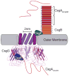
The curli biogenesis system possesses a unique outer membrane secretion apparatus. The CsgG pore is composed of eight subunits (the front three have been removed to improve clarity) with a 2nm wide central pore. Both CsgE and CsgF interact directly with the CsgG pore. CsgE is periplasmic and required for directing soluble CsgA to the CsgG pore for secretion while CsgF is surface exposed and contributes to the assembly of CsgB into a surface associated, amyloid-templating conformation. Once outside the cell, CsgA interacts with CsgB and assembles into an amyloid fiber.
Figure 3
Figure 3
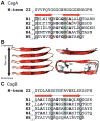
The molecular details of CsgA and CsgB. A. The mature CsgA protein is composed of an N-terminal 22 amino acids and 5 amyloid repeating units, each with a β-sheet-turn-β-sheet (indicated by the red arrows). Ser, Gln and Asn residues (boxes) in each repeating unit align in stacks that stabilize the amyloid conformation. The Gln residues at positions 49 and 139 and the Asn residues at positions 54 and 144 (circled) are essential for amyloid formation. Repeating unites R2, R3 and R4 contain “gatekeeper” residues (*) that temper amyloid formation. The conserved glycine and alanine residues are indicated in bold font. B. A cartoon representation of the predicted structure of CsgA shows the five repeating units assemble into a β-helical, cross-β structure (side view, left). The Gln, Asn and Ser residues of R1 (upper right) are shown as sticks and the overhead view of CsgA (lower right) shows the alignment of these residues within the predicted structure. C. The mature CsgB protein is composed of an N-terminal 22 amino acids and 5 amyloid repeating units with similarly conserved Gln and Asn stacks (boxes). The conserved glycine and alanine residues in R1-R4 are indicated in bold font. Repeating unit 5 lacks one of the Asn repeats, but instead contains four charged residues (indicated by +) that may be important for tethering CsgB to the cell surface via R5.
Figure 4
Figure 4
Curli subunit sharing between adjacent cells, or interbacerial complementation, is made possible by the nucleation-precipitation mechanism of curli fiber assembly. A csgB mutant (or csgA+ donor strain) secretes soluble CsgA into the extracellular milieu. CsgA from a csgB mutant can polymerize on a csgA mutant (or csgB+ acceptor) that is presenting CsgB on the cell surface when grown in close proximity to one another on a plate. The left panel shows a csgB mutant streaked near a csgA mutant, and on the edge of the CsgB-expressing streak there is a Congo red staining region.

References

    1. Hardy J, Selkoe DJ. The amyloid hypothesis of Alzheimer’s disease: progress and problems on the road to therapeutics. Science. 2002;297:353–356. - PubMed
    1. Breydo L, Wu JW, Uversky VN. Alpha-synuclein misfolding and Parkinson’s disease. Biochim Biophys Acta. 2012;1822:261–285. - PubMed
    1. Zuccato C, Valenza M, Cattaneo E. Molecular mechanisms and potential therapeutical targets in Huntington’s disease. Physiol Rev. 2010;90:905–981. - PubMed
    1. Chapman MR, Robinson LS, Pinkner JS, Roth R, Heuser J, Hammar M, Normark S, Hultgren SJ. Role of Escherichia coli curli operons in directing amyloid fiber formation. Science. 2002;295:851–855. - PMC - PubMed
    1. Romero D, Aguilar C, Losick R, Kolter R. Amyloid fibers provide structural integrity to Bacillus subtilis biofilms. Proc Natl Acad Sci U S A. 2010;107:2230–2234. - PMC - PubMed

Publication types

LinkOut - more resources