. 2013 Oct 17;5(1):207-15.
doi: 10.1016/j.celrep.2013.08.041. Epub 2013 Sep 27.

Human GEN1 and the SLX4-associated nucleases MUS81 and SLX1 are essential for the resolution of replication-induced Holliday junctions

Affiliations

Human GEN1 and the SLX4-associated nucleases MUS81 and SLX1 are essential for the resolution of replication-induced Holliday junctions

Elizabeth Garner et al. Cell Rep. .

Abstract

Holliday junctions (HJs), the DNA intermediates of homologous recombination, need to be faithfully processed in order to preserve genome integrity. In human cells, the BLM helicase complex promotes nonnucleolytic dissolution of double HJs. In vitro, HJs may be nucleolytically processed by MUS81-EME1, GEN1, and SLX4-SLX1. Here, we exploit human SLX4-null cells to examine the requirements for HJ resolution in vivo. Lack of BLM and SLX4 or GEN1 and SLX4 is synthetically lethal in the absence of exogenous DNA damage, and lethality is a consequence of dysfunctional mitosis proceeding in the presence of unprocessed HJs. Thus, GEN1 activity cannot be substituted for the SLX4-associated nucleases, and one of the HJ resolvase activities, either of those associated with SLX4 or with GEN1, is required for cell viability, even in the presence of BLM. In vivo HJ resolution depends on both SLX4-associated MUS81-EME1 and SLX1, suggesting that they are acting in concert in the context of SLX4.


Figures

Figure 1
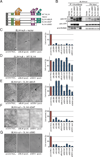
Figure 1. Depletion of BLM or GEN1 in the absence of SLX4 is synthetically lethal in human cells
(A). Schematic of SLX4 illustrating select domains and interacting nucleases, along with the N-terminally HA-tagged SLX4 cDNAs used in all experiments. Although the interaction of SLX1 and SLX4 has been shown to be direct, SLX4-XPF-ERCC1 and SLX4-MUS81-EME1 might not be direct. (B). Western analysis of immunoprecipitated HA-tagged SLX4 and co-immunoprecipitated XPF or MUS81 from cell lines used in the experiments that follow. The lower band (*) indicates degradation products. (C-G) Survival of SLX4 null cells complemented with indicated cDNAs and treated with siRNA against Luciferase (siCONTROL), siBLM, and siGEN1. SLX4 null cells complemented with empty vector (C), WT SLX4 cDNA (D), SLX4ΔMLR lacking interaction with XPF (E), SLX4ΔSAP lacking interaction with MUS81 (F), and SLX4ΔSBD that lacks interaction with SLX1 (G). Three separate siRNAs or a pool of three siRNAs were used for these studies as indicated. Left panels show cell morphology and the decreased cell number as observed using a phase contrast microscope. Right panels show cell survival as measured with Cell Titer-Glo. Survival data show average of at least six replicates for each siRNA and the error bars indicate standard deviations between replicates. Efficiency of BLM and GEN1 depletion is shown in Figure S1.
Figure 2
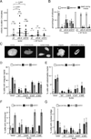
Figure 2. Mitotic duration is greatly increased and the majority of mitoses are abnormal in SLX4 deficient cells depleted of GEN1 or BLM
(A). Duration of mitosis (in minutes) assessed by live-cell imaging of GFP-H2B-positive SLX4-null cells complemented with vector or WT SLX4 and also transfected with the indicated siRNAs. Each value plotted represents a single cell undergoing mitosis. Cells were scored from 5 independent movies for each experimental condition. p values were determined by one-way ANOVA. (B). Outcome of mitoses in the experiment described in (A). Cells entering mitosis and segregating to yield two daughter cells were scored as ‘normal’. Cells that failed to segregate or did so with lagging chromosomes or multiple daughters were scored as ‘aberrant’. Cells that died during mitosis or those with daughter cells that died during mitotic exit were scored as ‘death during mitosis’. Values plotted represent the mean percentages of scores from 5 independent movies for each experimental condition. Error bars represent the standard deviation between the 5 replicates. See also Table S1. (C). to (G). Nuclear morphology of SLX4-null cells depleted of GEN1 or BLM. SLX4-null and complemented cells transfected with indicated siRNAs were stained with DAPI 96 hrs post second siRNA transfection. Nuclear morphology was scored according to the example panel shown in (C). Cells with irregular nuclei (D), with catastrophic nuclei (E), with micronuclei (F), and with nuclear bridges (G) were counted. Note that cells that die during mitosis were not counted since they were not attached to a coverslip. Values plotted represent the mean percentage from three experimental replicates. Error bars represent the standard deviation of the replicate means. See also Figure S2.
Figure 3
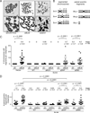
Figure 3. Depletion of BLM or GEN1 in the absence of SLX4 results in distinct chromosomal phenotypes that are dependent on MUS81 and SLX1
(A) Metaphase spreads from SLX4-null cells depleted of BLM or GEN1 showing presence of segmented chromosomes (siBLM) or paired acentric fragments (siGEN1). Upper panels display whole metaphases. The boxed area is shown below at higher magnification. Paired acentric fragments (arrowhead) appear to originate from telomere-proximal fragments that affect both sister chromatids (black arrow. Chromatid breaks are also seen (red arrows). See Figure S4 for staining for telomeric DNA. (B) Examples of scoring of sectors and paired acentric fragments used for quantification. (C) Quantification of segmented chromosomes in SLX4-null and complemented cells depleted of BLM. Chromosomes with 3 or more segments were scored in the indicated cell lines. There had to be at least two paired fragments, on the p and/or q arm, extending from the centric fragment, separated by gaps or breaks, for a total of at least three segments. SLX4-null cells depleted of GEN1 showed no segmented chromosomes. (D) The acentric fragments (distinct pairs only) were quantified in SLX4-null and complemented cells depleted of GEN1. 20 metaphases were scored for each experiment shown in this figure and the analysis was blinded. Means and standard deviations are shown above each graph. Significance was determined by one-way ANOVA. p values are indicated for the statistically significant comparisons of interest.
Figure 4
Figure 4. Exogenous expression of RusA rescues chromosomal segmentation or paired acentric fragmentation following depletion of BLM or GEN1 in SLX4-null cells
(A). SLX4-null cells were stably transduced with either empty vector, HA-tagged active RusA or HA-tagged inactive RusA (D70N). HA-tagged RusA expression levels are shown by anti-HA western analysis. (B). Quantification of segmented chromosomes in RusA expressing SLX4-null cells depleted of BLM. Chromosomes with 3 or more segments were scored in the indicated cell lines. (C). Quantification of paired acentric fragments in RusA expressing SLX4-null cells depleted of GEN1. Acentric fragments in distinct pairs only were scored in the indicated cell lines. (B) and (C) The number of metaphases scored, means and standard deviations are shown above each graph. Analysis was blinded for both experiments in which SLX4-null cells expressing vector control or active RusA were depleted of control or BLM protein. Significance was determined by one-way ANOVA. p values are indicated for the statistically significant comparisons of interest.
Figure 5
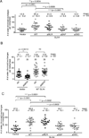
Figure 5. Both MUS81 and SLX1 interactions with SLX4 are necessary for the formation of SLX4-dependent sister chromatid exchanges
(A). Quantification of sister chromatid exchanges (SCEs) after SLX4-null and complemented cells were treated with low levels of interstrand crosslinking agents (one hour treatment with 0.1 micrograms of MMC per mL of media) (B). Quantification of SCEs after siRNA-mediated depletion of GEN1 from SLX4-null or WT complemented cells and treatment with low levels of interstrand crosslinking agents. (C). Quantification of SCEs after siRNA mediated depletion of BLM from SLX4-null and complemented cells. SCEs were scored blindly (A and C) and not blindly (B) on 20 metaphases from each cell line. Means and standard deviations are given above the graphs. Significance was determined by one-way ANOVA. p values are indicated for the statistically significant comparisons. See also Figure S4.
Figure 6
Figure 6. Model of Holliday junction processing in mitotically growing human cells
(1) The BLM-TOP3α-RMI1-RMI2 Holliday junction dissolvase complex facilitates branch migration of dHJs towards one another and decatenation of the DNA strands without the use of structure specific endonucleases. This process yields entirely non-crossover reaction products. (2) SLX4 associated nucleases provide essential nucleolytic processing of dHJs that are not dissolved in the absence of the BLM complex. The strict requirement of both MUS81 and SLX1 interaction with SLX4 for the suppression of synthetic lethality in the absence of BLM as well as the increase of SCEs after MMC treatment or BLM depletion suggests that they together form an in vivo HJ resolvase. (3) GEN1 is able to process HJs but is unable to prevent the synthetic lethality of SLX4-BLM deficiency. Both SLX4-dependent or GEN1 mediated resolution of dHJs may yield both cross-over and non-crossover products. A single Holliday junction or another substrate unsuitable for BLM complex mediated activity requires SLX4-complexed nucleases or GEN1. GEN1-SLX4 synthetic lethality indicates that GEN1 activity is necessary in the setting of SLX4 deficiency even in the presence of the BLM complex. In this setting, SLX4-interacting MUS81-EME1 and SLX1 are again required for the suppression of GEN1-SLX4 synthetic lethality.

Similar articles

Cited by

References

    1. Andersen SL, et al. Drosophila MUS312 and the vertebrate ortholog BTBD12 interact with DNA structure-specific endonucleases in DNA repair and recombination. Mol Cell. 2009;35:128–135. - PMC - PubMed
    1. Boddy MN, et al. Mus81-Eme1 are essential components of a Holliday junction resolvase. Cell. 2001;107:537–548. - PubMed
    1. Cejka P, et al. Rmi1 stimulates decatenation of double Holliday junctions during dissolution by Sgs1-Top3. Nat Struct Mol Biol. 2010;17:1377–1382. - PMC - PubMed
    1. Chaganti RS, et al. A manyfold increase in sister chromatid exchanges in Bloom's syndrome lymphocytes. Proc Natl Acad Sci U S A. 1974;71:4508–4512. - PMC - PubMed
    1. Chan SN, et al. Sequence specificity and biochemical characterization of the RusA Holliday junction resolvase of Escherichia coli. J Biol Chem. 1997;272:14873–14882. - PubMed

Publication types

MeSH terms