. 2013 Nov 8;113(11):1253-64.
doi: 10.1161/CIRCRESAHA.113.301787. Epub 2013 Sep 30.

Activation of NADPH oxidase 4 in the endoplasmic reticulum promotes cardiomyocyte autophagy and survival during energy stress through the protein kinase RNA-activated-like endoplasmic reticulum kinase/eukaryotic initiation factor 2α/activating transcription factor 4 pathway

Affiliations

Activation of NADPH oxidase 4 in the endoplasmic reticulum promotes cardiomyocyte autophagy and survival during energy stress through the protein kinase RNA-activated-like endoplasmic reticulum kinase/eukaryotic initiation factor 2α/activating transcription factor 4 pathway

Sebastiano Sciarretta et al. Circ Res. .

Abstract

Rationale: Autophagy is an essential survival mechanism during energy stress in the heart. Oxidative stress is activated by energy stress, but its role in mediating autophagy is poorly understood. NADPH oxidase (Nox) 4 is an enzyme that generates reactive oxygen species (ROS) at intracellular membranes. Whether Nox4 acts as a sensor of energy stress to mediate activation of autophagy is unknown.

Objective: We investigated whether Nox4 is involved in the regulation of autophagy and cell survival during energy stress in cardiomyocytes.

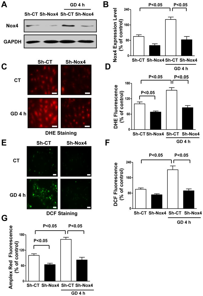
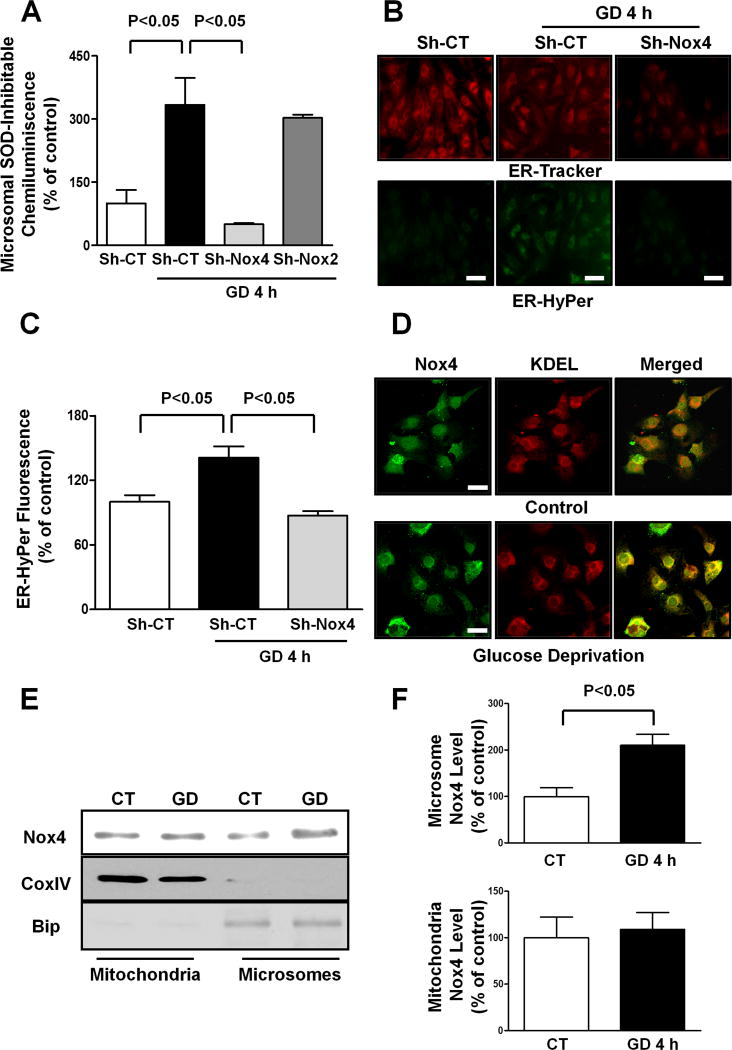
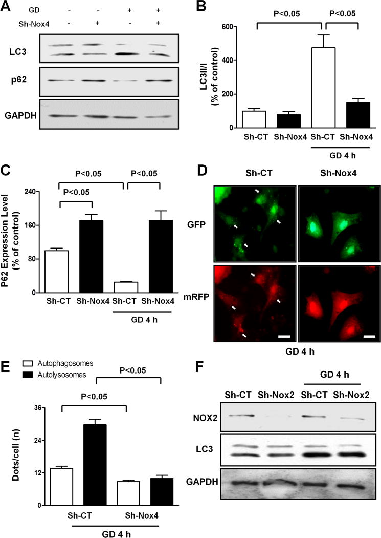
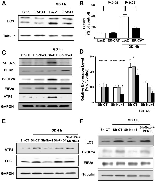
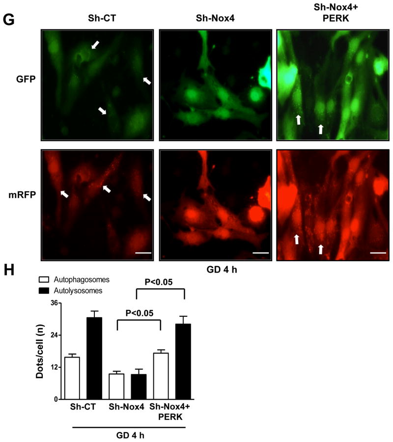
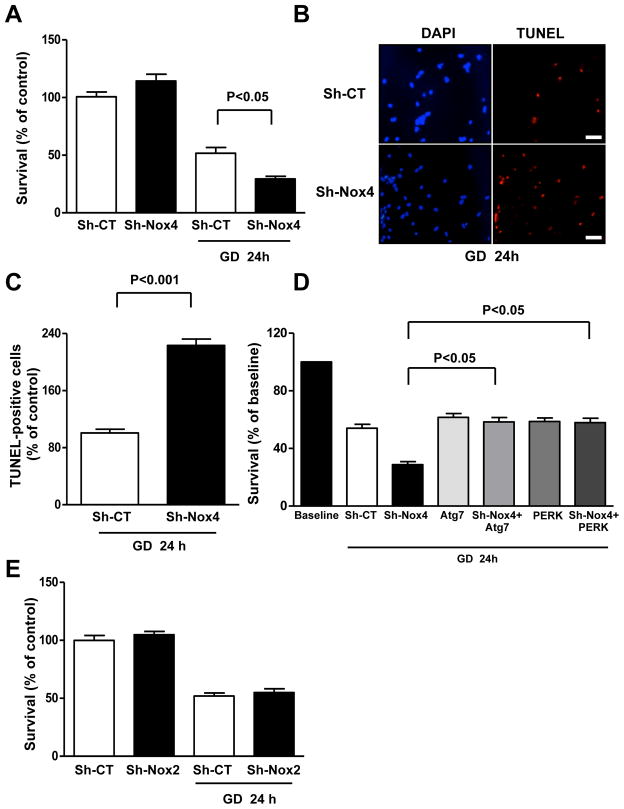
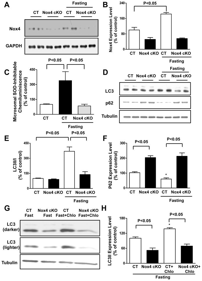
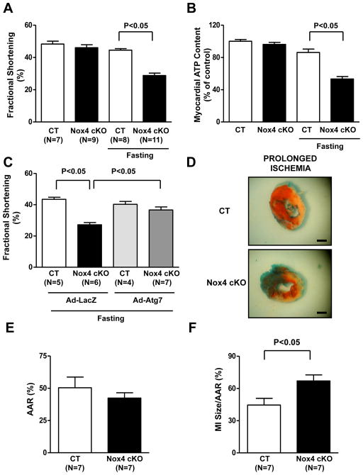
Methods and results: Production of ROS in cardiomyocytes was increased during glucose deprivation (GD) in a Nox4-dependent manner. Protein levels and the ROS-producing activity of Nox4 were increased in the endoplasmic reticulum (ER), but not in mitochondria, in response to GD. Selective knockdown of Nox4, but not Nox2, or selective reduction of ROS in the ER with ER-targeted catalase, but not mitochondria-targeted perioxiredoxin 3, abrogated GD-induced autophagy. Nox4 promoted autophagy during GD through activation of the protein kinase RNA-activated-like ER kinase pathway by suppression of prolyl hydroxylase 4. The decrease in cell survival during GD in the presence of Nox4 knockdown was rescued by reactivation of autophagy by Atg7 overexpression, indicating that the effect of Nox4 on cell survival is critically mediated through regulation of autophagy. Nox4 was activated during fasting and prolonged ischemia in the mouse heart, where Nox4 is also required for autophagy activation and cardioprotection.

Conclusions: Nox4 critically mediates autophagy in response to energy stress in cardiomyocytes by eliciting ROS in the ER and stimulating the protein kinase RNA-activated-like ER kinase signaling pathway.

Keywords: NOX4 protein; autophagy; endoplasmic reticulum; fasting; myocardial ischemia; reactive oxygen species.


Figures

Figure 1
Figure 1. Nox4 levels are increased in CMs during GD and promote ROS production
A–B. CMs were transduced with adenoviruses expressing a short hairpin sequence targeting Nox4 (Sh-Nox4) or short hairpin scramble (Sh-CT) for 96 hours and then subjected to GD. Densitometric analysis of Nox4 expression levels was performed. N=5. C–G. CMs with and without Nox4 knockdown were subjected to GD. DHE fluorescence (C–D), DCF fluorescence (E–F) and Amplex Red assay (G) were then performed to evaluate ROS levels. Data are presented as a percentage of control (Sh-CT). N=4–8. Bar=50 μm.
Figure 2
Figure 2. Nox4 is upregulated in the ER of glucose-deprived CMs
A. CMs were transduced with Sh-CT, Sh-Nox4 or Sh-Nox2 for 96 hours, and then CMs were cultured with normal or glucose-free medium for 4 hours. The microsomal fraction was isolated and a lucigenin assay to evaluate NADPH-dependent O2 production was performed. N=3–5. B–C. CMs were transduced with Sh-CT or Sh-Nox4 for 96 hours together with adenovirus expressing HyPer protein targeted to the ER (Ad-ER-HyPer) for the last 48 hours. CMs were cultured with normal or glucose-free medium for 4 hours. CMs were stained with ER-tracker red. Representative images of ER-Hyper and ER-tracker fluorescence are shown (B) together with quantification (C). Bar=50 μm. D. CMs were cultured with normal or glucose-free medium for 4 hours. An immunocytofluorescence assay of Nox4 and KDEL was performed and cells were observed with a confocal microscope. Bar=20 μm. E–F. In the same conditions, mitochondrial and microsomal fractions were isolated and Nox4 protein levels were assessed in each fraction. Representative immunoblots (E) together with densitometric quantification are shown (F). N=3. All values in the bar graphs are expressed as a percentage of the control.
Figure 3
Figure 3. Nox4 is required for autophagy activation in CMs during GD
A–C. CMs were transduced with Sh-CT or Sh-Nox4 for 96 hours, and then CMs were cultured with normal or glucose-free medium. LC3 and p62 protein levels were evaluated. Representative immunoblots (A) and densitometric quantification (B–C) are shown. N=4. Data are presented as a percentage of Sh-CT. D–E. CMs were transduced with Sh-CT or Sh-Nox4 for 96 hours together with Ad-mRFP-GFP-LC3 for the last 48 hours. CMs were cultured with glucose-free medium. Representative images of mRFP and GFP dots are shown (D), together with quantification of autophagosomes and autolysosomes (E). N=5. Bar=10 μm. F. CMs were transduced with Sh-CT or Sh-Nox2 for 96 hours, and then CMs were cultured with normal or glucose-free medium. LC3II and Nox2 levels were evaluated.
Figure 4
Figure 4. Nox4 activation in the ER promotes autophagy in energy-deprived CMs through activation of the PERK/eIF-2α/ATF4 pathway
A–B. CMs were transduced with Ad-LacZ or Ad-ER-catalase for 48 hours, and then CMs were cultured with normal or glucose-free medium for 4 hours. LC3II levels were evaluated. Representative immunoblots (A) and densitometric quantification (B) are shown. C–D. CMs were transduced with Sh-CT or Sh-Nox4 for 96 hours, and then CMs were cultured with normal or glucosefree medium. Phospho-PERK (Thr980), phospho-eIF-2α (Ser51) and ATF4 protein levels were evaluated. Representative immunoblots (C) and densitometric quantification (D) are shown. N=4. # p<0.05 vs. Sh-CT; * p<0.05 vs. Sh-CT and Sh-Nox4 GD. E. CMs were transduced with Sh-CT or Sh-Nox4 for 96 hours, with and without Sh-PHD4. CMs were cultured with normal or glucose-free medium. ATF4 and LC3 levels were evaluated. F. CMs were transduced with Sh-CT or Sh-Nox4 for 96 hours, together with Ad-LacZ or Ad-PERK for the last 48 hours. CMs were cultured with normal or glucose-free medium. LC3II and phospho-eIF-2α levels were evaluated. G–H. CMs were transduced with Sh-CT or Sh-Nox4 for 96 hours, together with Ad-LacZ or Ad-PERK and with Ad-mRFP-GFP-LC3 for the last 48 hours. CMs were cultured with glucose-free medium. Representative images of mRFP and GFP dots are shown (G), together with quantification of autophagosomes and autolysosomes (H). N=4. Bar=10 μm.
Figure 5
Figure 5. Nox4 disruption reduces CM survival and promotes apoptosis in CMs during GD
A–C. CMs were transduced with Sh-CT or Sh-Nox4 for 96 hours, and then CMs were cultured with normal or glucose-free medium for 24 hours. Cell survival was assessed by Cell Titer Blue assay (CTB, A). N=4. Percentage of TUNEL-positive cells was also calculated. Representative pictures (B), together with quantification (C) are presented. N=3. Bar=50 μm D. CMs were transduced with Sh-CT or Sh-Nox4 for 96 hours, together with Ad-LacZ or Ad-Atg7 or Ad-PERK for 48 hours. CMs were then cultured with normal or glucose-free medium. Cell survival was assessed by CTB. N=4. Data are expressed as percentage of baseline (cells cultured with regular medium). E. CMs were transduced with Sh-CT or Sh-Nox2 for 96 hours, and then CMs were cultured with normal or glucose-free medium. Cell survival was then assessed by CTB. N=3.
Figure 6
Figure 6. Nox4 is activated and promotes autophagy in the heart during fasting in vivo
A–B. Control and Nox4 cKO mice were subjected to 48 hours of fasting. Cardiac Nox4 protein abundance was evaluated. Representative immunoblot (A) and densitometric analyses are shown (B). N=4. C. Control and Nox4 cKO mice were subjected to 48 hours of fasting. Cardiac microsomes were isolated and NADPH-dependent O2 production was assessed by lucigenin assay. N=4–6. D–F. Control and Nox4 cKO mice were subjected to 48 hours of fasting. Cardiac LC3 and p62 protein levels were evaluated. Representative immunoblots (D) and densitometric analyses are shown (E–F). N=4–5. G–H. Control and Nox4 cKO mice were subjected to 48 hours of fasting, and some received chloroquine (10 mg/kg i.p) 4 hours before they were sacrificed. Representative immunoblot of cardiac LC3 is shown, together with quantification analysis. N=3. * p<0.05 vs. CT. Data are expressed as a percentage of the control.
Figure 7
Figure 7. Nox4 disruption in the heart is detrimental during energy deprivation in vivo
A. Control and Nox4 cKO mice were subjected to 48 hours of fasting and then underwent echocardiographic evaluation. Fractional shortening was calculated. B. Control and Nox4 cKO mice were subjected to 48 hours of fasting. Myocardial ATP content was evaluated. N=4. Data are expressed as a percentage of the control. C. Control and Nox4 cKO mice received cardiac injection of Ad-LacZ or Ad-Atg7. After 48 hours, they were subjected to 48 hours of fasting. Fractional shortening was calculated. D–F. Control and Nox4 cKO mice were subjected to 3 hours of ischemia. Representative pictures of 2,3,5-triphenyltetrazolium chloride staining are shown (D), together with quantification of the area at risk (AAR, E) and myocardial infarct size (MI/AAR, F). Bar= 1 mm.

References

    1. Singh R, Cuervo AM. Autophagy in the cellular energetic balance. Cell Metab. 2011;13:495–504. - PMC - PubMed
    1. Sciarretta S, Zhai P, Shao D, Maejima Y, Robbins J, Volpe M, Condorelli G, Sadoshima J. Rheb is a critical regulator of autophagy during myocardial ischemia: pathophysiological implications in obesity and metabolic syndrome. Circulation. 2012;125:1134–1146. - PMC - PubMed
    1. Hariharan N, Maejima Y, Nakae J, Paik J, Depinho RA, Sadoshima J. Deacetylation of FoxO by Sirt1 Plays an Essential Role in Mediating Starvation-Induced Autophagy in Cardiac Myocytes. Circ Res. 2010;107:1470–1482. - PMC - PubMed
    1. Kuma A, Hatano M, Matsui M, Yamamoto A, Nakaya H, Yoshimori T, Ohsumi Y, Tokuhisa T, Mizushima N. The role of autophagy during the early neonatal starvation period. Nature. 2004;432:1032–1036. - PubMed
    1. Matsui Y, Takagi H, Qu X, Abdellatif M, Sakoda H, Asano T, Levine B, Sadoshima J. Distinct roles of autophagy in the heart during ischemia and reperfusion: roles of AMP-activated protein kinase and Beclin 1 in mediating autophagy. Circ Res. 2007;100:914–922. - PubMed

Publication types

MeSH terms

LinkOut - more resources