

Case Reports
doi: 10.1007/s12105-013-0494-4.
Epub 2013 Oct 1.
Clear cell cystic variant of calcifying epithelial odontogenic tumor
Affiliations
- PMID: 24081908
- PMCID: PMC4022929
- DOI: 10.1007/s12105-013-0494-4
Item in Clipboard
Case Reports
Clear cell cystic variant of calcifying epithelial odontogenic tumor
Head Neck Pathol.
2014 Jun.
Abstract
Calcifying epithelial odontogenic tumor (CEOT) is a solid, locally aggressive, benign odontogenic neoplasm characterized by sheets and nests of polyhedral epithelial cells exhibiting eosinophilic and less often clear cytoplasm, occasional nuclear pleomorphism without mitotic activity, calcifications, and deposits of amyloid. A cystic variant has been reported only twice. Herein, we present an additional example of cystic CEOT occurring in a 31-year-old male and featuring clear cell epithelial lining with deposits of amyloid and osteodentin.
Figures

a Orthopantomogram. Unilocular mixed radiopaque/lucent, expansile and locally aggressive lesion, located in the left posterior mandible and ascending ramus; b CT. Osteolytic lesion is seen with expansion of surrounding tissue and a central area isodense to bone

Gross features. Portion 1 features cystic cavity with projections into the lumen. Portion 2 features calcified spherical and irregular mass
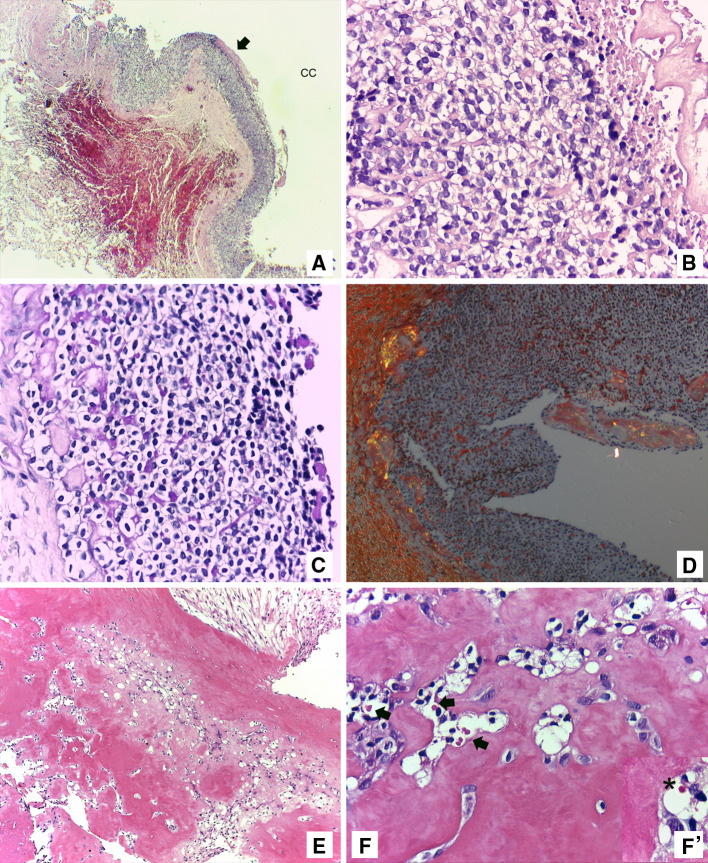
a Cystic cavity (CC) lined by odontogenic epithelium of generally uniform thickness, with focal necrosis (arrow) (H&E ×40); b Clear cells in the cystic lining revealing intracellular and eosinophilic extracellular material (H&E ×400); c Positive diastase resistant deposits (PAS. diastase stain ×400); d Amorphous, eosinophilic hyalinized material exhibiting apple-green birefringence after polarization (Congo red stain ×100); e Calcified material with characteristics of osteodentin (H&E ×100); f Round eosinophilic globules consistent with “thanatosomes” (arrows) (H&E ×400) and f’ “thanatosome” in detail (asterisk) (H&E ×1,000)
References
-
- Pindborg JJ, Simonsen EK, Hansen PB. A case of calcifying epithelial odontogenic tumor. Tandlaegebladet. 1966;70:822–830. - PubMed
-
- Mesquita RA, Lotufo MA, Sugaya NN, De Araújo NS, De Araújo VC. Peripheral clear cell variant of calcifying epithelial odontogenic tumor: report of a case and immunohistochemical investigation. Oral Surg Oral Med Oral Pathol Oral Radiol Endod. 2003;95:198–204. doi: 10.1067/moe.2003.63. - DOI - PubMed
Publication types
MeSH terms
Supplementary concepts
LinkOut - more resources
Full Text Sources
Other Literature Sources
Medical
