. 2013 Nov 15;191(10):5196-203.
doi: 10.4049/jimmunol.1300354. Epub 2013 Sep 30.

Lipopolysaccharide stimulates platelets through an IL-1β autocrine loop

Affiliations

Lipopolysaccharide stimulates platelets through an IL-1β autocrine loop

G Thomas Brown et al. J Immunol. .

Abstract

LPS activates platelets through TLR4, aiding productive sepsis, with stimulated splicing and translation of stored heteronuclear pro-IL-1β RNA. Although the IL-1R type 1 (IL-1R1) receptor for IL-1 shares downstream components with the TLR4 receptor, platelets are not known to express IL-1R1, nor are they known to respond to this cytokine. We show by flow cytometry and Western blotting that platelets express IL-1R1, and that IL-1β and IL-1α stimulate heteronuclear I-1β splicing and translation of the newly made mRNA in platelets. Platelets also respond to the IL-1β they make, which is exclusively associated with shed microparticles. Specific blockade of IL-1R1 with IL-1R antagonist suppressed platelet stimulation by IL-1, so IL-1β stimulates its own synthesis in an autocrine signaling loop. Strikingly, IL-1R antagonist inhibition, pharmacologic or genetic suppression of pro-IL-1β processing to active cytokine by caspase-1, or blockade of de novo protein synthesis also blocked LPS-induced IL-1β mRNA production. Robust stimulation of platelets by LPS therefore also required IL-1β amplification. Activated platelets made IL-1β in vivo as IL-1β rapidly accumulated in occluded murine carotid arteries by posttranscriptional RNA splicing unique to platelets. We conclude that IL-1β is a platelet agonist, that IL-1β acts through an autocrine stimulatory loop, that an IL-1β autocrine loop is required to amplify platelet activation by LPS, and that platelets immobilized in occlusive thrombi are activated over time to produce IL-1β. IL-1 is a new platelet agonist that promotes its own synthesis, connecting thrombosis with immunity.


Figures
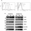
Figure 1
Figure 1. Human platelets express IL-1 receptors
A. Flow cytometric analysis of platelet surface type I IL1 receptors. Freshly isolated human platelets were stimulated with LPS, CD14, and LPS binding protein for 1h (LPS) or left unactivated (Ctrl) and then fixed with formaldehyde before incubation with polyclonal goat anti-IL1 type I receptor antibody (IL1R1) or with goat isotype control antibody (II°) followed by Alexa Flour488-conjugated anti-goat. Some platelets were left unstained before analysis of fluorescent events in a predetermined platelet gate. n=3. B. Platelets express the type I IL1 receptor protein. Increasing amounts (25, 50 75 μg) of protein from freshly isolated human platelet (left) and peripheral blood monocyte (right) lysates were resolved by SDS-PAGE before IL1R1, monocyte CD14, platelet CD42b, and common HSP90 detected by immunostaining as described in “Methods” to form a composite western blot. Representative of three experiments.
Figure 2
Figure 2. IL-1 receptor agonists activate human platelets
A. Soluble IL-1α and IL-1β stimulate platelet pro-IL-1β hnRNA splicing. Washed human platelets were treated with buffer or recombinant IL-1β (100 pg/ml), recombinant IL-1α (100 pg/ml), or E. coli LPS/recombinant CD14/recombinant LPS-binding protein (100 ng/ml each) for 3 h before total RNA was collected and analyzed for spliced pro-IL-1β mRNA by quantitative reverse transcriptase PCR. Shaded bars depict the effect of pre-incubation (30 min) with the IL1 specific IL1 receptor inhibitor IL1Ra (150 ng/ml) prior to incubation with the stated agonists. n= 5. B. Platelet stimulation by IL-1β requires MyD88. Human platelets purified (2 × AutoMACS) free of contaminating monocytes were treated with buffer or recombinant IL-1β (100 pg/ml, 3h) with or without pre-incubation with IL1Ra (150 ng/ml for 30 min) or an inhibitory MyD88-antennapedia chimeric peptide (50 μg/ml, 30 min). Cellular RNA was analyzed for accumulation of spliced pro-IL-1β mRNA by qRT-PCR as described in methods. N=3. Significance attained using One-way ANOVA *P-Value <0.05.
Figure 3
Figure 3. Platelet stimulation by LPS or LPS-derived microparticles requires IL1R1 signaling
A. Microparticles from LPS-stimulated platelets stimulate naïve platelets through IL1R1. Microparticles from LPS-treated (μLPS) or buffer-treated (μCtrl) platelets were recovered and washed by centrifugation before addition to naïve purified platelets before spliced pro-IL-1β mRNA levels were quantified 3 h later by qPCR. n=3. Significance attained using One-way ANOVA *P-Value <0.05 for the shown comparison and from control cells. B. IL1Ra blockade of IL1R1 prevents platelet stimulation by LPS. Washed human platelets were treated with buffer, recombinant IL-1β (100 pg/ml), the combination of phenol-extracted E. coli LPS (100 ng/ml) along with recombinant CD14 (100 ng/ml) and LPS-binding protein (100 ng/ml), or a stimulatory TRAF6 ligand-antennapedia chimeric peptide (5 μg/mL) for 3 h before total RNA was collected and analyzed for spliced pro-IL-1β mRNA by qRT-PCR. Some cells were exposed to the inhibitor IL1Ra at 150 ng/ml for 30 min before stimulation. n= 5.
Figure 4
Figure 4. LPS platelet stimulation requires IL-1β production
A. LPS induction of pro-IL-1β hnRNA splicing requires de novo protein synthesis. Purified platelets were treated for 3 h with LPS, recombinant CD14, and recombinant LPB (100 ng/ml each) in the presence or absence of cycloheximide (CHX, 100 μg/ml) prior to quantifying spliced pro-IL-1β RNA by qPCR. n=3. *< 0.05 for the shown comparison. B. Caspase inhibition blocks platelet LPS stimulation. Purified platelets were treated for 3 h with LPS, recombinant CD14, and recombinant LPB (100 ng/ml each, 3 h) in the absence or after 30 min pre-incubation with caspase-1 inhibitors (z-WEHD-fmk or z-YVAD-fmk, 100 μM) before spliced pro-IL-1β RNA was quantified. n=3 *< 0.05 for the shown comparison. C. IL-1 β hnRNA splicing in murine platelets requires caspase-1. Platelets of wild-type or caspase-1−/− animals were stimulated with LPS along with LPS-binding protein and soluble CD14 (100 ng/ml each) for 3h, treated with 100 pg/ml human IL-1β, or left unstimulated for 3h before spliced pro-IL-1β mRNA was quantitated by qPCR. n=2. Significance attained using One-way ANOVA *< 0.05 for the shown comparison, and from control cells.
Figure 5
Figure 5. IL-1β accumulates in platelets during thrombus maturation
A. IL-1β accumulates within arterial thrombi over time. Occlusive thrombi were induced by a brief (1 min) ectopic application of 7.5% FeCl3 to carotid arteries followed by sacrifice at the stated times as described in “Methods.” Fixed arterial sections were stained with hematoxylin and probed with rabbit non-immune or rabbit anti-mouse IL-1β (1:250) antibody. Primary antibodies were detected with HRP-conjugated anti-rabbit secondary antibody and visualized through formation of brown peroxidase reaction product. B. Thrombus IL-1β associates with anuclear platelet deposition. Thrombi were induced in carotid arteries as above and harvested at the stated times, fixed and sectioned before staining platelet membranes with Alexa594-conjugated wheat germ agglutinin (red), IL-1β with rabbit anti-mouse IL-1β and Alexa488-conjugated goat anti-rabbit (green), or nuclei with DAPI (blue). The control was isotype non-immune rabbit antibody. Sections were imaged (63×) by confocal microscopy, the images pseudocolored and overlaid.
Fig. 6
Fig. 6. Accumulation of thrombus IL-1β is independent of transcription
Mice were injected with actinomycin D 30 min prior to initiation of arterial injury by application of 7.5% FeCl3 to an exposed carotid artery. Anesthetized mice with surgical windows maintained at 37° were sacrificed 3h later, arterial segments fixed with OTC and sections stained with anti-P-selectin detected with Alexa Flur488-secondary antibody that fluoresces in the green channel (top left) and anti-IL1β detected with Alexa Flur568-secondary antibody that fluoresces in the red channel (top right) as nuclei were stained with DAPI mounting media that produces blue nuclear fluorescence (bottom right).

References

    1. Dinarello CA. Immunological and inflammatory functions of the interleukin-1 family. Annu. Rev. Immunol. 2009;27:519–550. - PubMed
    1. Dinarello CA. Interleukin-1beta and the autoinflammatory diseases. N. Engl. J. Med. 2009;360:2467–2470. - PubMed
    1. Qu Y, Franchi L, Nunez G, Dubyak GR. Nonclassical IL-1 beta secretion stimulated by P2X7 receptors is dependent on inflammasome activation and correlated with exosome release in murine macrophages. J. Immunol. 2007;179:1913–1925. - PubMed
    1. Andrei C, Dazzi C, Lotti L, Torrisi MR, Chimini G, Rubartelli A. The secretory route of the leaderless protein interleukin 1beta involves exocytosis of endolysosome-related vesicles. Mol. Biol. Cell. 1999;10:1463–1475. - PMC - PubMed
    1. MacKenzie A, Wilson HL, Kiss-Toth E, Dower SK, North RA, Surprenant A. Rapid secretion of interleukin-1beta by microvesicle shedding. Immunity. 2001;15:825–835. - PubMed

Publication types

MeSH terms

LinkOut - more resources